CT
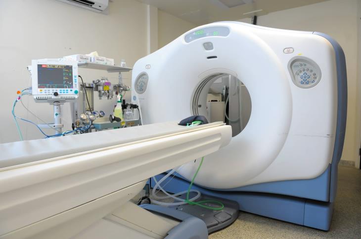
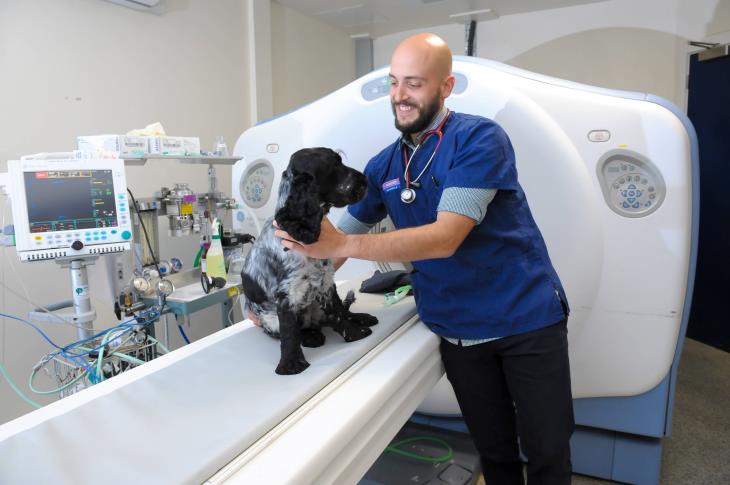
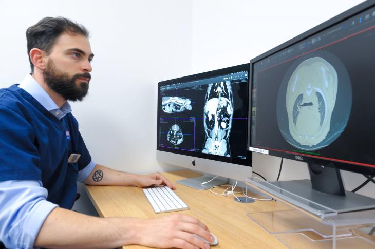
The CT room is home to an advanced 64 slice GE CT scanner which produces detailed cross sectional images allowing precise lesion identification. Using software we can reconfigure these into a detailed three-dimensional images which helps in visualising lesions and fractures and planning surgical treatment.
CT images are useful for assessing all kinds of diseases including bone abnormalities, spinal problems, teeth problems, nasal disease and conditions of the lungs. CT also helps us to assess patients with cancer since we can see much more detail about the extent of the tumour and whether it has spread elsewhere in the body. CT is especially useful to createhighly detailed images of lung tissue allowing the detection of lesions that are not visible on conventional radiographs. CT is also used for assessment of the blood vessels and anatomical abnormalities.
Radiographic contrast agents are often used, to help highlight areas of inflammation or abnormal tissue.


