Categories
An unusual presentation of corneal sequestrum in an American Bulldog
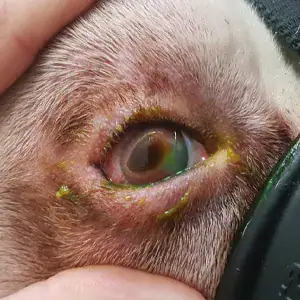
Obie an American Bulldog was referred to Specialist-led Ophthalmology team following a 10 day history of a dark pigmented corneal ulceration in the right eye and evidence of inwards rolling of the eyelids (entropion) causing the eyelashes and haired skin to rub on the surface of the eye (trichiasis).
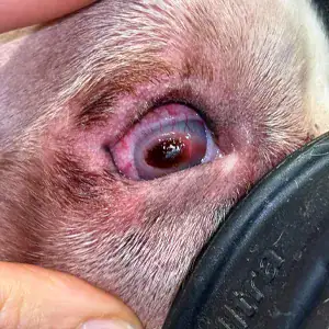
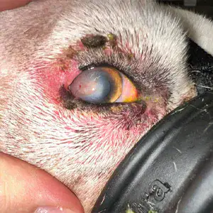
On examination, the eye was uncomfortable with marked to severe conjunctival hyperaemia (redness), blepharospasm (squinting) and discharge. The vision in the right eye was affected by the changes to the cornea, but there were still light reflexes indicating that the eye was still functioning beneath all of the surface pathology. The large brown plaque (corneal sequestrum) lateral to the centre of the cornea was associated with a bed of granulation tissue and well-established corneal blood vessels.
There was inwards rolling of the lower eyelid (entropion) that was causing the lashes and hair from the eyelid to rub against the cornea (trichiasis) in the location of the corneal changes. The upper lid also had some entropion but milder than the lower and not causing trichiasis.
Tear production and pressure were within normal limits.
Obie was admitted to Eastcott Referrals and surgery was performed under general anaesthetic to remove the sequestrum and correct the underlying eyelid abnormalities responsible.
Corneal surgery is very delicate (as the cornea in a healthy dog eye is less than 1mm thick) and is carried out at Eastcott under the high magnification of a surgical microscope.
The sequestrum was cut from the cornea using a crescent knife (a special scalpel with a flat blade for separating the layers of the cornea) leaving a bed of granulation with no traces of pigment. The eyelids were shortened to make them a little tighter which was sufficient to correct the upper eyelid entropion. A Hotz Celcus procedure, to remove some of the skin below the eyelid margin and roll the eyelid into a normal position along with shortening was used to correct the entropion of the lower eyelid.
After surgery Obie was managed with topical antibiotics and (due to peri-ocular dermatitis) systemic antibiotics, pain relief and topical application of donor plasma to reduce the risk of further loss of corneal depth.
With the exception of dermatitis around the eye and wound causing a continuation of the antibiotics, Obies recovery was uneventful. The sequestrum was sent for histopathology examination at an external laboratory to confirm the diagnosis.
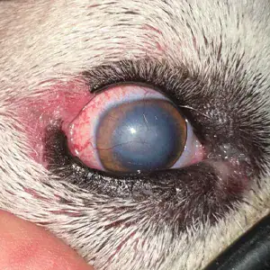
At the last examination Obie was comfortable with reasonable corneal clarity, which we would expect to continue to clear over time.
Corneal sequestrum is commonly seen in cats with chronic inflammation of the cornea (keratitis) due to entropion or viral infection but is unusual to see in a dog. The sequestrum often progresses deeper into the cornea making excision more complicated. Surgical excision is often combined with a corneal graft to provide support and protection if the cornea is significantly weakened by the reduction in thickness.
Due to the unusual presentation of Obie’s case he has been included in a research project being carried out by our colleagues in the Ophthalmology team at Willows Veterinary Centre and Referral Service.