Categories
Anterior Lens Luxation in Dogs
Anterior lens luxation (ALL) can occur as a result of a primary inherited weakness in the attachments of the lens or as a secondary consequence of another intraocular disease (e.g. uveitis, hyper mature cataract and glaucoma). Primary ALL is frequently seen in middle aged terrier breeds (especially the Jack Russell) and the Border Collie.
Four cases of anterior lens luxation
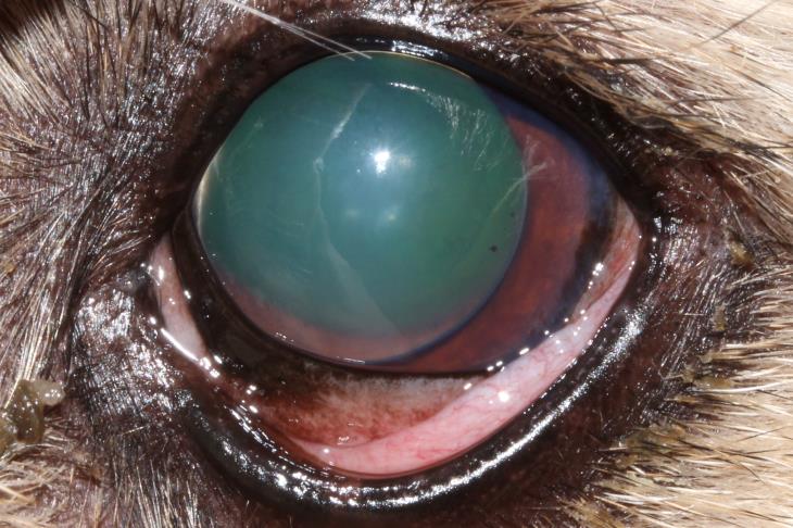
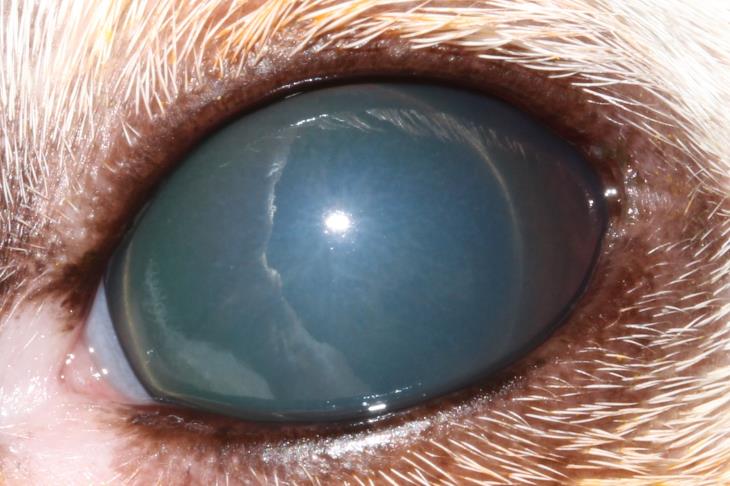
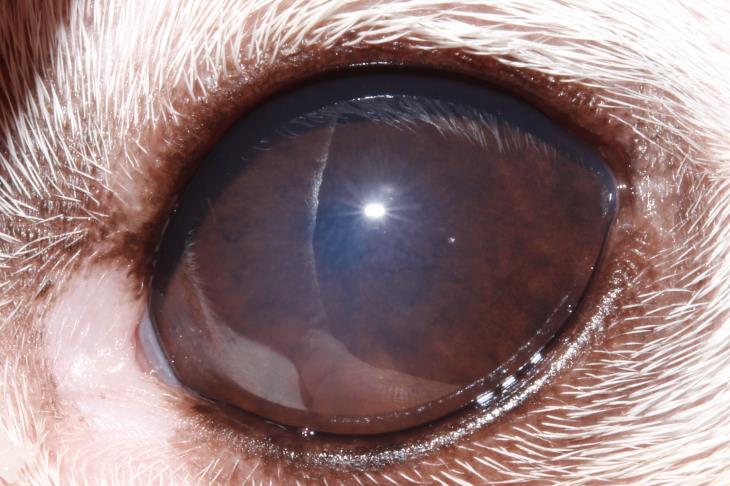
The crystalline lens can be seen in the anterior chamber in front of the iris
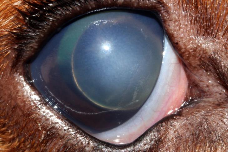
Case 1. ALL secondary to glaucoma in a Bassett Hound FN 8yrs.
Case 2. Primary ALL in a Jack Russell Terrier MN 6yrs.
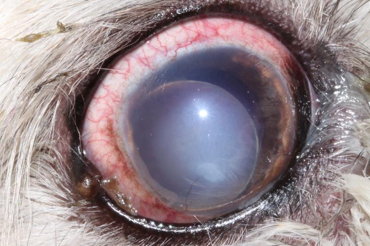
Case 3. ALL secondary to chronic uveitis in a Maltese Terrier FN 10 yrs.
Case 4. Primary ALL in a Jack Russell Terrier FN 4yrs.
Diagnosing ALL in Dogs
It can sometimes be difficult to identify ALL with the naked eye. Clinical signs associated with ALL include a raised or erratic intraocular pressure, scleral injection, a focal corneal oedema (this is the result of the lens touching the corneal endothelium) and of course a lens in the anterior chamber. Taking a photograph (with flash) can be a great help as the edge of the lens will often be clearly highlighted. In cases of ALL the whole of the pupillary margin will not be clearly visible as it will be at least partially obscured by the lens. If it is still unclear or if dense corneal oedema makes visually identifying ALL impossible then an ultrasound examination can be very helpful.
Treatment options for ALL in Dogs
Treatment options can be divided into the surgical removal of the lens or medical management following the reduction of the lens into the posterior chamber (‘couching’). Surgical removal of the lens can be achieved via a large corneal incision and the extraction of the intact lens inside its capsule (intracapsular lens extraction (ICLE)) or via a small incision using phacoemulsification to break up the lens inside the eye before aspirating it in a similar fashion to cataract removal. ‘Couching’ involves applying careful pressure through the cornea in order to reduce the lens back behind the iris and then trapping in there by inducing miosis. ‘Couching’ has been recently studied and found to have comparable success rates to surgical intervention. The procedure can be performed under sedation or conscious in a co-operative patient. This is not a viable option if the lens has formed adhesions to the iris (synechiae) and may be less successful if ALL has been present for more than two weeks.
Case 4 following couching of the lens and medical therapy to induce miosis. The eye is comfortable and visual
ALL in Dogs Summary
- Suspect ALL in any case of unexplained corneal oedema
- Terrier breeds especially the Jack Russell are most commonly effected
- ALL most commonly occurs between 3 and 7 years of age
- Treatment can be medical or surgical
- Closely monitor the fellow eye
 Mark is happy to discuss any Ophthalmology cases that you may have.
Refer a Case to Mark Ames or the Ophthalmology Team