Categories
Babesia in Small Animals
Babesia canis in Dermacentor reticulatus ticks
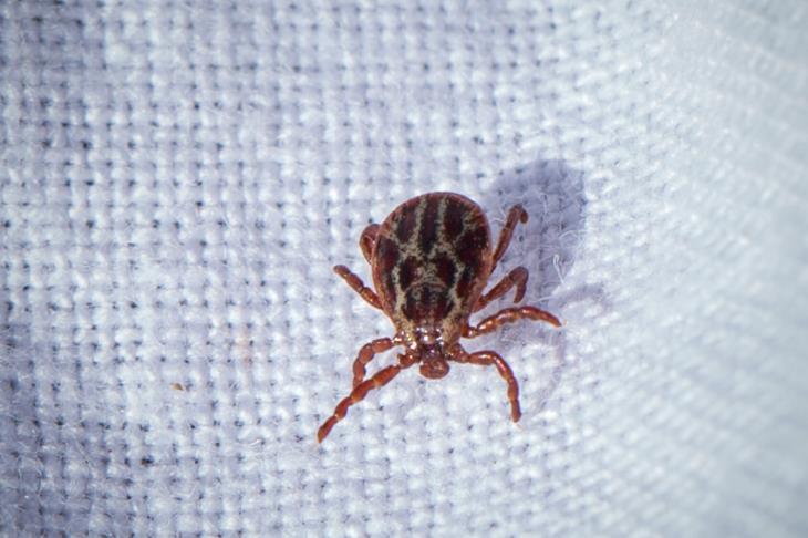
Following identification of Babesia canis in Dermacentor reticulatus ticks in Essex earlier this year and an associated small cluster of clinical babesiosis cases in untraveled dogs, internal medicine specialist Jenny Reeve reviews the presenting signs, diagnosis and management of babesiosis.
Babesia spp. are intraerythrocytic protozoan parasites, predominantly tick transmitted, although in the absence of vector presence, transmission is reported secondary to dog fights, blood transfusions and transplacental transfer. Large Babesia spp. include B. canis, B. rossi and B. vogeli and small Babesia spp. include B. gibsoni and B. conradae.
The spectrum of disease associated with Babesia spp. ranges from subclinical infections through to life threatening sequelae depending upon both parasitic factors (infecting spp. and strain, with B. rossi considered particularly virulent) and host factors (predominantly immunocompetence). Haemolysis arises secondary to immunologic and non-immunologic removal of both parasitised and uninfected red blood cells; thus the severity of disease is not directly related to parasite burden. Associated alterations in erythrocyte membranes and activation of inflammatory cascades may result in erythrocyte sludging within capillary beds and be responsible for some of the reported complications of the disease, including the central nervous system signs seen in some patients. Thrombocytopenia is commonly encountered, sometimes in the absence of anaemia, and may be due to secondary immune- mediated destruction or consumptive processes.
Examination findings are reflective of haemolytic anaemia (pallor, tachycardia, bounding pulse quality, haemoglobinaemia/uria, icterus, splenomegaly, pyrexia), the systemic inflammatory response and specific complications associated with individual cases.
Laboratory Findings
Laboratory findings include anaemia, whilst this is typically strongly regenerative, in acute disease this may appear non-regenerative (due to the 3-5 day delay in regenerative response), thus the possibility of babesiosis should not be excluded based upon an initial non-regenerative anaemia. As a result of the immunological basis for haemolysis, spherocytosis may be observed and both saline agglutination and Coombs’ testing may be positive. Thrombocytopenia is a common concurrent finding although clinical bleeding associated with this is rare. Serum biochemistry findings are non-specific and reflective of degree of haemolysis (haemoglobinaemia, hyperbilirubinaemia), hypoperfusion/ hypoxia (increased hepatic enzyme activities, hyperlactataemia) and specific complications (e.g. hypoglycaemia, azotemia; uncommonly observed).
Definitive diagnosis of Babesia spp. infection
- Blood film examination; this generally has poor sensitivity (high chance of false negative results). Whilst ampling from a peripheral capillary bed (e.g. ear tip) may increase sensitivity, it remains too poor for this to be an adequate screening test. Visualisation of typical intra-erythrocytic parasites enables identification of large and small Babesia spp. but does not differentiate between subspecies.
- Serology (immunoflourescent antibody; IFA); a positive result demonstrates exposure to Babesia spp. but this may be historic and does not confirm Babesia spp. are the definitive cause of the current presenting signs. False negative results may be seen in early disease due to the delay in serologic response (repeating convalescent serology may be required), very young dogs or occasionally, inexplicably, in chronically infected dogs. Serology cannot be used for speciation as cross-reactivity occurs between Babesia spp.
- PCR (blood, less commonly splenic aspirates); has the greatest sensitivity for diagnosis in acute infection and a positive result confirms active infection. It is the only practical test for reliable differentiation between subspecies; this is of clinical importance as therapy differs between species. Initially a general Babesia spp. PCR (to screen for all Babesia spp.) with subsequent subspecies specific PCR (to identify the subspecies) is suggested. The subspecies PCRs are highly specific and will fail to identify other subspecies and are therefore inappropriate screening tests.
Clinical syndromes and signs that may be associated with Babesia spp. Infection
- Haemolytic anaemia +/- associated haemoglobinaemia/uria, icterus
- Thrombocytopenia
- Splenomegaly
- Non-specific pyrexia, inappetance, lethargy, weakness
- Less commonly: neurological complications, glomerulonephritis and/or acute kidney injury, respiratory distress, rhabdomyolysis systemic inflammatory response and disseminated intravascular coagulation
Treatment
A variety of treatment protocols (off licence use in the UK) have been described although imidocarb dipropionate is the preferred therapy for large Babesia spp. and combination therapy with atovaquone/azithromycin is most effective for small Babesia spp. The aim of therapy in nonendemic regions should be clearance of the organism; PCR 60 and 90 days after therapy may be used to evaluate success, which is not always achieved. Adjunctive treatments such as fluid therapy and blood product administration may be required in some cases. Occasionally the severity of concurrent immune-mediated
haemolysis complicates treatment and if anti-protozoal therapy alone is not effective, in some cases a concurrent rapidly tapering glucocorticoid course may be of benefit.
Prevention of Babesia spp. infection
Preventative measures include avoiding travel of dogs to endemic regions, prophylactic tick control (transmission may take a minimum of 2 days following attachment), vaccination (generally considered to offer some protection to B. canis and B. rossi and limit severity rather than incidence of disease), minimizing aggressive interaction between dogs and appropriate testing of blood donors and products to avoid iatrogenic transmission. In regions of geographic prevalence, the possibility of subclinical babesiosis should be considered prior to either splenectomy or chemotherapy; both of which are risk factors for subsequent development of clinical babesiosis.
Babesia spp. are prevalent in warmer geographic regions and remain rare in untraveled UK dogs. However, with recent demonstration of presence within competent vectors and progressive climate change, frequency of clinical babesiosis is likely to increase and should be considered as a differential diagnosis in dogs with appropriate presenting signs.
Feline babesiosis
Feline babesiosis is considered less prevalent and therefore less well studied than the canine counterpart. A variety of feline specific, large and small, Babesia spp. of varying pathogenicity have been found to affect cats. Feline babesiosis typically follows a more chronic course and as a result of this, severe, clinically compensated, regenerative anaemia is often identified. Treatment of feline babesiosis is significantly more complicated as many anti-babesial drugs described in dogs are not effective and of those with reported efficacy, there is little margin between therapeutic and lethal drug doses.