Categories
Canine Ear Disease – Quiz
What is your diagnosis?

with Tim Charlesworth
History
“Fencer” is a 9 year old Male (neutered) WHWT with a 12 month history of intermittent bilateral otitis externa. Fencer has had a left sided head tilt and also yelps with pain when yawning and refuses to eat his biscuits. Initial investigations are unrewarding with little response to NSAID therapy and topical ear medication based on culture/sensitivity. Fencer is anaesthetised and a CT examination performed (see below)
‘
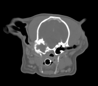
CT image
What are the most significant findings?
Click for answer
What is the most likely diagnosis in this case?
Click for answer
What is the treatment of choice?
Click for answer
Read Summary….
Tim is hosting a free CPD evening on 4th March 2015
Ear Surgery in Dogs – How, When & Why?
Places are still available Book a place…
Answer:
The left tympanic bulla is distorted and irregularly enlarged with areas of irregular bony proliferation and also osteolysis including erosion of the medial bulla wall towards the cranial vault. Some localised meningeal enhancement is visible suggestive of a meningitis. >>back to quiz
Answer
The most likely diagnosis is Cholesteatoma which is becoming increasingly frequently diagnosed. Other ddx would include a slowly growing primary middle ear neoplasm or otitis media (less likely).
Cholesteatoma is an epidermoid cyst that forms within the middle ear cavity. Contrary to what the name suggests, it is non-neoplastic and has nothing to do with cholesterol or fat. Rather, it is charaterised by a cornifying stratified squamous epithelium which produces keratin leading to a slowly expansile lesion within the tympanic bulla. The aetiology is incompletely understood but, although congenital forms are possible, they are most likely a result of chronic otitis externa in which keratinizing stratified epithelium has had the opportunity to seed and establish within the middle ear chamber.
Although in theory the cyst is benign, it can cause a range of signs relating to its position and size. Symptoms therefore include signs of otitis media – head tilt, nystagmus, circling etc but animals can also present as dogs who are reluctant to fully open their mouths and/or are reluctant to eat and this is due to encroachment of the cholesteatoma on the temperomandibular joint (TMJ). We have also seen some dogs which presented with dysphagia due to orpharyngeal compression from ventral outgrowth from the bulla. The lesions can also extend medially and erosion of the cranial vault can lead to otitis interna and even meningoencephalitis. >>back to quiz
Answer
The treatment of choice is surgical removal of the keratin contents together with the secretory lining responsible for the lesion and this involves either total ear canal ablation and lateral bulla osteotomy or ventral bulla osteotomy (or both!). Surgery can be challenging as the distorted anatomy makes it hard to remove all of the responsible epithelium and this leads to recurrence which is reported in approximately 50% of cases (Hardie et al 2008). Recurrence is most likely in dogs with either neurological signs, bone lysis or inability to open the mouth.
Summary
With the increasing availability of advanced imaging (CT, MRI etc) increasing numbers of Cholesteatoma are being diagnosed and so it is being regarded as an emerging disease. There is no reason to think that this is the case, however with cholesteatoma recognised in 10% of cases of otitis media in the early 1990’s (Little et al 1991). We are now better at diagnosing the condition, however, and early detection of these lesions leads to better clinical outcomes. We therefore advise considering CT examination of dogs with persistent otitis externa and early surgical intervention in dogs which are diagnosed with this condition. >>back to quiz