Categories
CT Visualisation of Deep Vegetal Foreign Bodies
Soft Tissue Referral Team take advantage of the Diagnostic Imaging Department for the visualisation of deep vegetal foreign bodies using CT
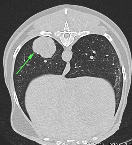
Otto the Cocker Spaniel was referred to us following a persistent cough of 3 months duration. Antibiotics and anti-inflammatory treatment had been prescribed by the referring vet and when there had been no improvement after 2 weeks of treatment, a possible deep bronchial foreign body was suspected. Chest x-rays had been taken and radiographic findings were consistent with a focal area of increased radiopacity in the right caudal pulmonary lobe. An endoscopy had then been performed and two grass seeds were found in the main right caudal bronchus, 2cm distally from the tracheal bifurcation.The presence of another large foreign body in the deeper portion of the affected bronchus was suspected. However, due to the inaccessibility of the area, Otto was referred to us for a thoracic CT scan in order to clearly assess the suspicious lobe. Computed tomography multi-planar reconstruction (MPR) and 3D reconstruction of the lungs highlighted the presence of a 3 cm foreign body in the main right bronchus of the caudal pulmonary lobe, approx. 8 cm beyond the tracheal bifurcation. An endoscopy was subsequently performed and the team was able to remove an ear of corn, along with numerous detached seeds and associated debris that had trapped in the lung for many weeks. Otto was discharged the following day and no further signs of coughing have been reported by the owner.
body with soft tissue density (green arrowheads).
There is a mild amount of gas bubbles trapped by the foreign body.
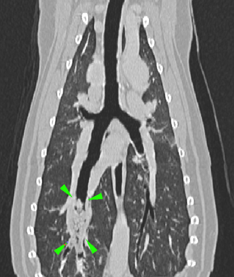
bronchus is partially occluded by a foreign body with
soft tissue density (green arrowheads). There is a mild
amount of gas bubbles trapped by the foreign body
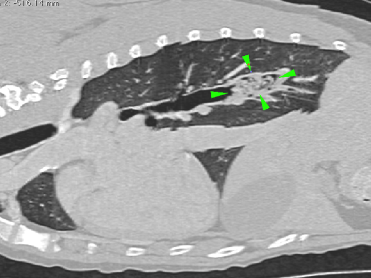
right caudal
bronchus is partially occluded by a foreign body
with soft tissue density (green arrowheads).
There is a mild amount
of gas bubbles trapped by the foreign body
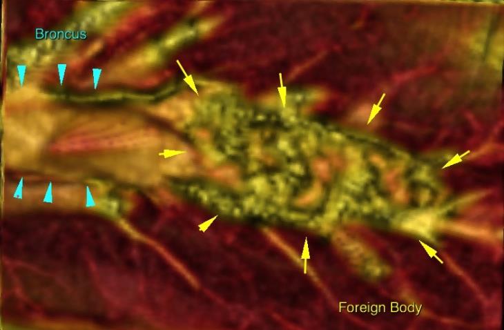
caudal bronchus(light blue arrowheads),
with the foreign body trapped in it (yellow
arrows).
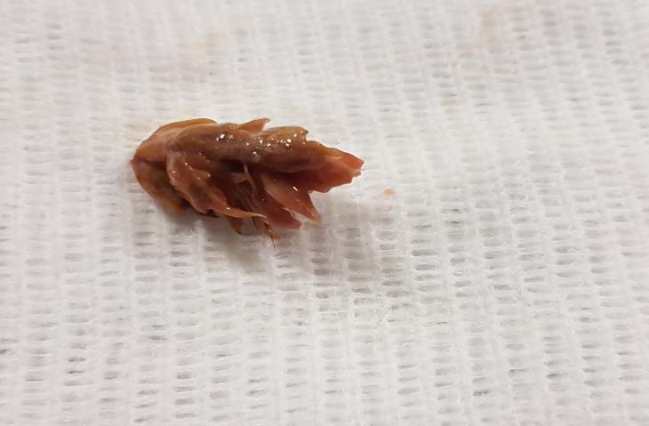
from the affected bronchus with
the help of the bronchoscope.
Read blog on Ultrasound aided retrieval of vegetal foreign bodies
 with Domenico Sainato DMV, MRCVS, MSc (Small Animal Diagnostic Imaging)
If you would like to refer a case for investigation, please refer via the Soft Tissue Referral Service