Categories
Eastcott Referrals Dentistry Case Studies
5 small animal dentistry case studies from the Eastcott Referrals dentistry team
- 1. Repairing a bilateral fractures to the rostral mandible
- 2. Treating a benign but invasive oral tumour
- 3. Collaboration on custom made Titanium plates
- 4. An oro-nasal fistula
- 5. Pulp exposure in a canine tooth fracture
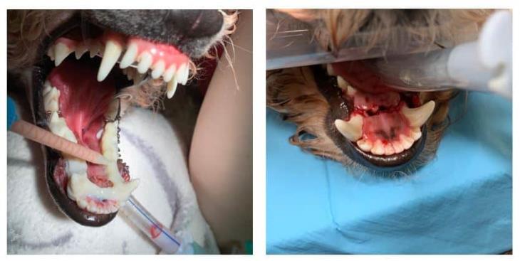
1. Repairing a bilateral fractures to the rostral mandible
After a collision with a park bench, this patient suffered bilateral fractures to the rostral mandible and both mandibular canine teeth were dislocated. In order to preserve the patient’s teeth and maintain a normal occlusion, the canine teeth were repositioned and a wire and acrylic splint applied.
The soft tissue lacerations were also sutured. A transmylohyoid intubation was performed to aid access and monitor the occlusion during the placement of the splint. This will be re-evaluated in 4 weeks to check healing and determine if the canine teeth are still vital. Luxated/ avulsed teeth must receive re-implantation treatment in less than 72 hours to have a good success rate of continuing to be vital teeth.
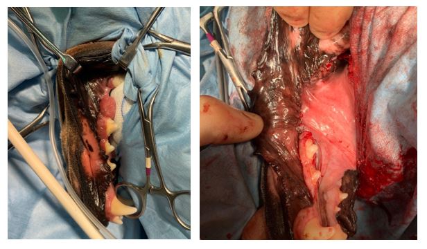
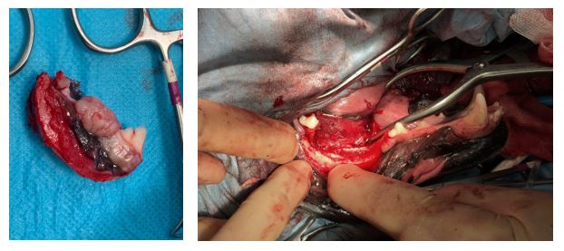
2. Treating a benign but invasive oral tumour
These are pictures of a rim resection of a mandible for a acanthomatous ameloblastoma (a benign but invasive oral tumour) in a dog. This type of resection is conservative in its approach, biomechanically secure in its design and maintains normal occlusion. It can be a very good option for treating less aggressive or invasive forms of tumours affecting the mandible in medium and large breed dogs.
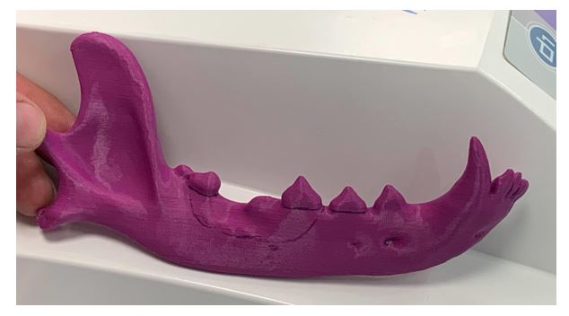
3. Collaboration on custom made Titanium plates
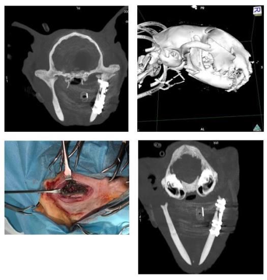
We’ve been collaborating with James Sage and Dan Jones from Fusion Implants and Ben Walton from ChesterGates Veterinary Specialists to design a range of 3D printed custom Titanium plates for caudal mandibular fractures in cats.
Caudal mandibular fractures in cats are challenging because the ramus is usually less than 2.5cm tall, in some areas only 1.5mm thick and access to the surgical site is limited.
We used a plate for the first case last week on a vertical fracture of the right mandibular ramus. The plate adapted to the mandible and helped to reduce the fracture very well and our patient was eating the day after surgery.
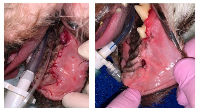
4. An oro-nasal fistula
Communication between the nose and the mouth, an oro-nasal fistula, can develop due to numerous pathological processes. They are more common in breeds with longer noses, as in this Daschund. Extraction of a periodontally diseased tooth and subsequent local infection has resulted in this patient’s fistula.
Food material and saliva can enter the nose causing long term irritation and pain. Successful repair of these defects is technique sensitive. This patient was treated by a buccal mucosal advancement flap, closed in 2 layers, effectively closing the communication.
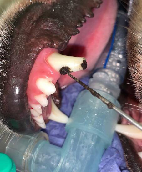
5. Pulp exposure in a canine tooth fracture
Here we have a one month old complicated crown fracture of a canine tooth. This fracture involves enamel, dentin and exposes the sensitive pulp.
Pulp exposure is both painful and opens the pulp to bacteria. Pulpitis and eventually, pulp necrosis will have occurred as a result of this trauma.
Endodontic treatment was performed to remove the black necrotic pulp and enable the patient to keep this strategic tooth. Without treatment these cases can develop into potentially destructive abscesses.