Categories
Eddie – a minimally invasive cardiac surgery case
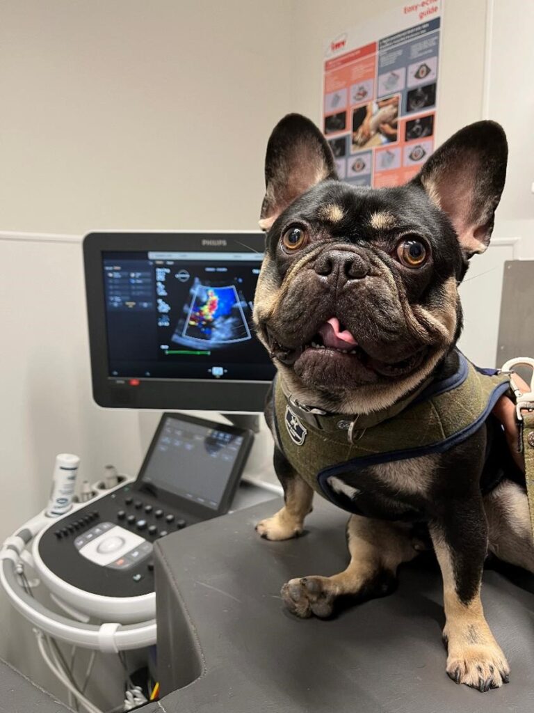
Eddie, a French bulldog, was diagnosed with severe pulmonary valve stenosis (PS) after investigation of episodes of fainting whenever he was excited. He was diagnosed with early heart failure, and prescribed medication to reduce fluid accumulation.
His heart scan showed an over-loaded right side of the heart, and an under-filled left heart. Severity of PS was graded as severe (125 mmHg, normally below 30 mmHg). Pulmonary valve stenosis can be successfully managed by performing a minimally invasive surgical procedure, called a balloon valvuloplasty. Here, fluoroscopic imaging is used to guide the placement of cardiac catheters and wires across the narrowed pulmonary valve, and a balloon dilation catheter is inflated to open the fused valve. After removal of the catheters and wires, only a small surgical wound remains. This requires very little post-operative pain relief, and minimal disruption to routine.
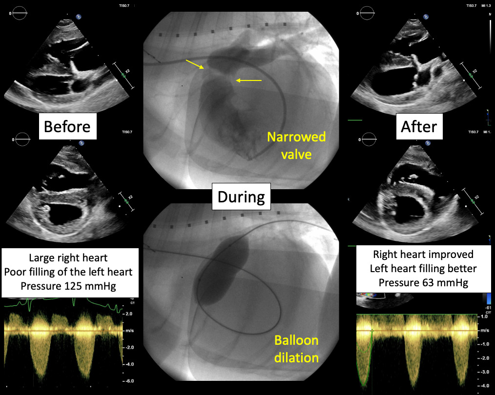
Eddie’s PD procedure went very well. At his 4-week re-evaluation, we identified that the pressure on his heart had reduced by half, and the left side of his heart was much better filled. His fainting has resolved, and he no longer requires treatment for heart failure.
We will continue to monitor Eddie annually, but his quality of life is significantly improved after his minimally invasive cardiac surgery and he seems very happy with the result!