Categories
Gingivitis Stomatitis Complex in Cats
Overview
Gingivitis stomatitis complex (GSC) is believed to be an inappropriate response to normal plaque and viral antigens found in the oral cavity. The complete pathogenesis is not fully understood, however there is an inflammatory response which is cellular in nature with plasmacytic -lymphocytic infiltration of the mucosal and gingival tissues. The trigger for the development of GSC has not been identified but the vast majority of individuals (in some publications 100%) are infected with calici virus. Retro-viral infection has also been linked to GSC but this is not a commonly identified co-infection in this clinic.
All of the oral mucosal surfaces may be affected with severe hyperaemia, ulceration and proliferation. There is commonly secondary periodontitis and tooth resorption but for a diagnosis of GSC there must be extension of pathology beyond the muco-gingival junction. The inflammatory changes are usually widespread, although focal lesions are less commonly identified. The inflammation is often more severe toward the caudal oral cavity, especially immediately adjacent to the maxillary and mandibular molar teeth and the caudal oral mucosa (often incorrectly referred to as the fauces).
There have been studies suggesting a greater prevalence in pedigree cats although the majority seen at this clinic are domestic short hair cats which are likely to reflect the population that we serve. No sex predilection has been identified and a widespread age range is reported, we commonly see patients as young as 12 months at time of initial presentation.
Diagnosis
Tentative diagnosis can be proposed on clinical appearance and duration but routine biochemistry, haematology, urine analysis and Calici PCR are appropriate screening tests with definitive diagnosis based on biopsy, which is especially important for asymmetric or focal lesions. The lack of response to simple periodontal treatment or the mismatch between the severity of inflammation and the levels of plaque and calculus are also important.
Patients may present with a highly variable clinical picture, from apparently asymptomatic through to grossly debilitated with foul halitosis, anorexia and continuous ptyalism. It can be challenging to highlight the potential severity of the disease to owners when cats are not exhibiting extensive symptoms but the disease is commonly progressive and early intervention is apparently associated with an improved prognosis.
Treatment & Management
Plaque control is at the heart of effective management of these cases. Chronic ulcerative paradontal stomatitis, seen in dogs, is believed to have a similar pathogenesis and these patients can often be effectively managed with rigorous home care and regular periodontal prophylactic therapies. For the majority of cat owners this is not a viable option. Plaque is a highly stable biofilm and the bacteria within it live in a highly privileged environment effectively secluded from parenteral antibiotics and therefore any improvement of the pathology with antibiotic therapy is likely to be short lived and may be counter productive with the development of resistant populations. Anti-inflammatory medication, steroidal or non-steroidal, may be of benefit if welfare is grossly affected but will not effect plaque levels and will not prevent progression of periodontitis. To date, elective extraction has been the management technique of choice with the highest rate of success in clinical trials.
Elective Extractions
Elective extractions have been defined as complete and confirmed extraction of all teeth that are encircled by the inflamed mucosa or are affected by another existing pathology. In practice this can mean extraction of all of the teeth or more commonly the pre-molar and molar teeth only. There has not been a statistical difference between full and partial mouth extractions identified. Understandably clients are concerned by the prospect of such surgery and how it may affect the patient and we are always careful to provide appropriate counseling. Cats are extraordinarily adaptable and more reliant on their tongue to prehend prepared cat diets than their teeth. Although it is likely cats may not be able to chew dry foods effectively this commonly does not prevent them from trying.
Approximately half of the patients managed by elective extraction show a complete response with resolution of all symptoms and clinical signs. A further third of patients improve dramatically but may need long term medical management to augment the benefits of surgery. Adjunctive medical management usually comprises non-steroidal anti-inflamatories and recombinant interferon omega given orally. All of the adjunctive therapies have proven less efficacious if used without elective extraction. Some have suggested that the use of essential fatty acids, anti-oxidants and stress management may be beneficial but as yet evidence is not available. Use of cyclosporine for refractory cases may be effective and adjunctive pain management, especially targeted toward chronic or neuropathic pain, can be employed in the short or long term.
Incomplete extraction of teeth has been a cause of poor response to treatment and so pre and post extraction dental radiographs are exceedingly important. Extraction technique, with focus on minimizing surgical trauma, has a significant role in achieving short term comfort and post operative patient recovery.
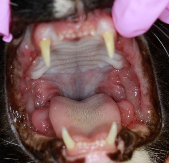
1. Photograph of cat with Gingivitis-Stomatitis Complex showing severe inflammation of gingival and non gingival oral tissue with proliferation of the mucosa in the caudal oral cavity
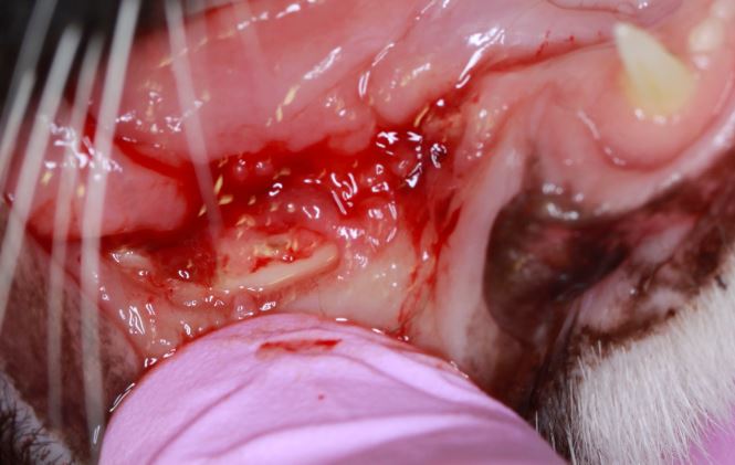
2. Photograph of the right mandible of a cat with Gingivitis-Stomatitis Complex showing full thickness mucosal ulceration at the site of the incomplete extraction of the distal root of the mandibular fourth pre-molar
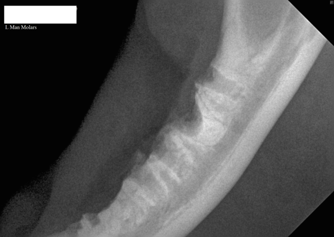
3. Intra-oral radiograph of the left mandible of a cat with a retained mesial root of the molar tooth
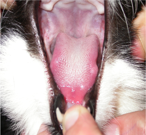
4. Photograph of the oral cavity of a cat with complete resolution of the signs of GSC following extraction of all premolar and molar teeth

Both Peter Southerden and Andrew Perry are happy to discuss or give advice on all matters relating to small animal veterinary dentistry or oral surgery. Please phone 01793 528341 or email Eastcott Referrals