Categories
How Computed Tomography can help us in the diagnosis of diseases of the head
What’s your diagnosis…?
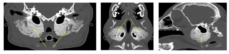
A 6-month-old West Highland White Terrier was referred to our hospital due to severe oral pain and the impossibility to open his mouth. A CT scan was performed and a severe, bilateral and symmetric ‘palisading’ periosteal reaction, was observed at the level of the temporal bones (yellow arrows). The temporomandibular joints and the tympanic bullae were within normal limits.
 Scroll down to reveal the diagnosis……
 Scroll down to reveal the diagnosis……
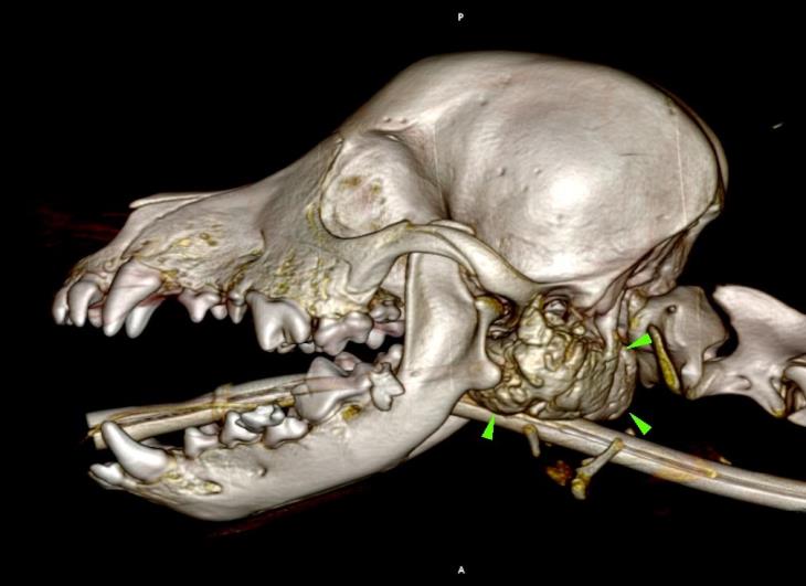
A diagnosis of craniomandibular osteopathy was made; an autosomal recessive, self-limiting developmental disease, primarily seen in Highland terriers.
A 3D reconstruction of the head is also displayed below, with the lesion highlighted by green arrowheads.
The primary use of CT imaging is of the head of dogs and cats with neurological or nasal diseases. Technologic advances in hardware and software, opened up new possibilities, including abdominal and thoracic imaging, and high definition scanning of lungs and bones.
CT is now widely used in veterinary practice and has become an essential asset to veterinarians in the pursuit of many diagnoses. Long body parts can be scanned within a couple of seconds in amazing detail and accurate contrast resolution.
Thanks to the possibility of reformatting the images in any plane, or as three-dimensional projections, a better representation of the structural anatomic relationships can be achieved.
CT’s diagnostic power is comparable with, or superior to, other imaging modalities such as ultrasound and MRI for many disorders and has immense potential as a rapid and efficient diagnostic tool for a wide range of indications.