Categories
Peri-apical bone loss in the left maxillary premolar tooth
History
The dog was presented with a swelling on the left side of his face. The patient is a two year old male Springer Spaniel.
222
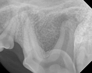
x-ray of the dog’s maxillary premolar
Questions
- Which tooth is this?
- What pathology can you see?
- What is the most likely cause?
- What are the treatment options for this tooth?
Answers
- This is the left maxillary fourth premolar tooth (sometimes referred to as a carnassial tooth). Dogs typically have four maxillary and four mandibular premolars. The maxillary fourth premolar is a three rooted tooth with two mesial and one distal roots
- There is evidence of peri-apical bone loss affecting all three roots of the left maxillary premolar tooth. This will be associated with inflammation and often infection in this area
- The most likely cause is a complicated crown fracture. Complicated crown fractures are tooth fractures that expose the pulp. Pulp exposure will result in infection and inevitable pulp necrosis
- The treatment options for this tooth are either extraction or endodontic treatment. Teeth that have pulp exposure should always be treated. Extraction of teeth in dogs is a surgical procedure which often involves raising a muco-periosteal flap, removing bone and sectioning the tooth into single rooted sections. It is a significant surgical procedure with potential complications. Endodontic treatment would involve root canal treatment in this case. The benefits of endodontic treatment are that it is non-invasive, there are fewer potential complications and an important strategic tooth is preserved