Categories
Pericardiectomy in Small Animals
Pericardiectomy is one of the most common therapeutic thoracoscopic procedures that is performed in veterinary patients. Pericardiectomy is indicated in patients that have pericardial effusion that restricts diastolic filling causing cardiac tamponade and associated abdominal effusion. Pericardial effusion can be caused by neoplasia (e.g. haemangiosarcoma, chemodectoma) in which case pericardiectomy is palliative. Most commonly, however, pericardial effusions are judged to be idiopathic and pericardiectomy can be curative in these cases. All cases require full echocardiography prior to surgery in order to pre-operatively identify any possible mass lesions that may require biopsy or removal or affect the expected prognosis.
Thoracoscopic sub-total pericardiectomy/pericardial window techniques can be safely performed using 3, 5-10mm incisions. The required section of pericardium is removed and submitted for histology and a chest drain placed to allow evacuation of air from the thorax and monitoring/removal of any postoperative effusion. The majority of dogs are discharged the next day once the drain has been removed. The long-term prognosis (for cases of idiopathic effusion) is excellent.

Figure 1: the thickened pericardium has been entered and a suction probe is used to remove any residual pericardial effusion
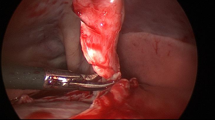
Figure 2 & Video Clip: a patch of pericardium is resected and submitted for histopathology
We are more than happy to discuss any possible referrals with our referring vets including assessing whether or not MIS approaches are suitable for a particular approach. Please do contact us if you have any queries or want to find out more.