Categories
The use of a novel orthopaedic plate in the treatment of a fracture
The use of a novel orthopaedic plate in the treatment of a fracture of the lateral part of the right humeral condyle
– by Eastcott Referrals Orthopaedic Surgeon Fabio Frazzica
An 8-year-old male Cocker Spaniel was presented to Eastcott Referrals after having been diagnosed with a traumatic fracture of his right humerus by his local vets.
His radiographs had shown a displaced fracture of the lateral part of the right humeral condyle.
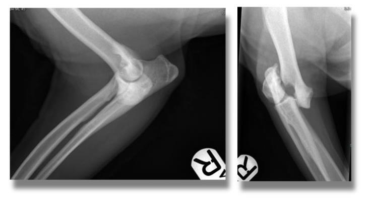
This represents a fracture involving the elbow joint and it requires careful surgical repair in order to restore limb function and prevent the onset of severe osteoarthritis.
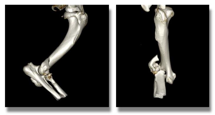
A CT scan of the affected limb was obtained to allow for preoperative planning and to rule out additional fissures that might have gone undetected on plain radiography.
In order to obtain a strong and reliable repair, internal fixation with a transcondylar screw and an orthopaedic plate and screws was chosen.
Up until recently, the standard way of treating such fractures would have involved the use of plates that are contoured to the patient’s bone shape at the time of surgery. This can be a time-consuming procedure which prolongs the duration of the anaesthetic. Furthermore, bending a metal plate to the desired shape increases the risk of implant failure due to the formation of micro-fissures within the metal itself.
To overcome these limitations, a team of Orthopaedic Specialists and Biomechanicals at Fusion Implants has developed an implant that precisely replicates the shape of the surface of the lateral part of the humeral condyle and thus does not require additional contouring in most cases.
Eastcott Referrals is proud to have been chosen as one of the first few centres in the UK to use this novel implant known as the Lateral Epicondylar Anatomical Plate (LEAP).
This patient underwent surgery and made a good recovery from his general anaesthetic.
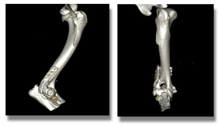
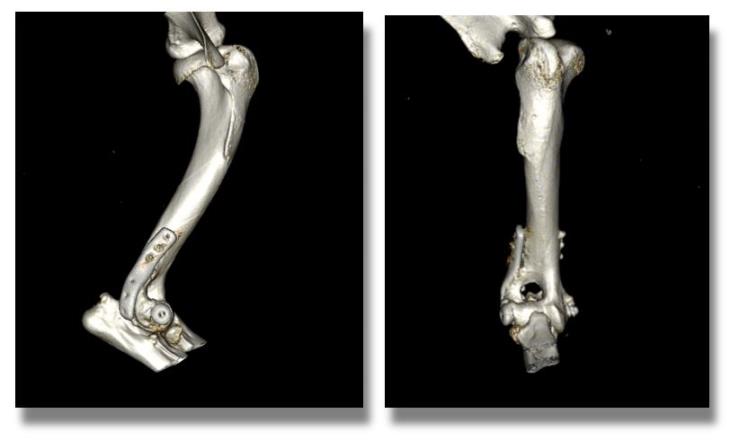
His postoperative CT scans below show excellent apposition and alignment of the bone fragments and restoration of a normal joint surface.
Two months later the patient a further CT scan was performed to assess his progress and we were pleased to confirm excellent osseous union and no implant-associated complications.
He is currently doing well enjoying an active lifestyle and has not required long-term medications nor exercise restriction.