Categories
What’s your diagnosis? Computed Tomography Case 3
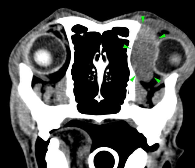
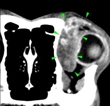
A 7-years-old Labrador was presented to with lethargy, anorexia and exophthalmos.
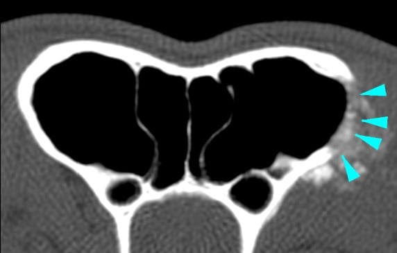
A CT scan of the head revealed the presence of a soft tissue space-occupying lesion of approximately 3 cm in diameter, arising from the orbital part of the left frontal bone (green arrowheads). The mass was laterally compressing the ipsilateral eye. A focal osteolytic process was observed at the level of the left zygomatic process of the frontal bone (light blue arrowhead).
Scroll down for the diagnosis…
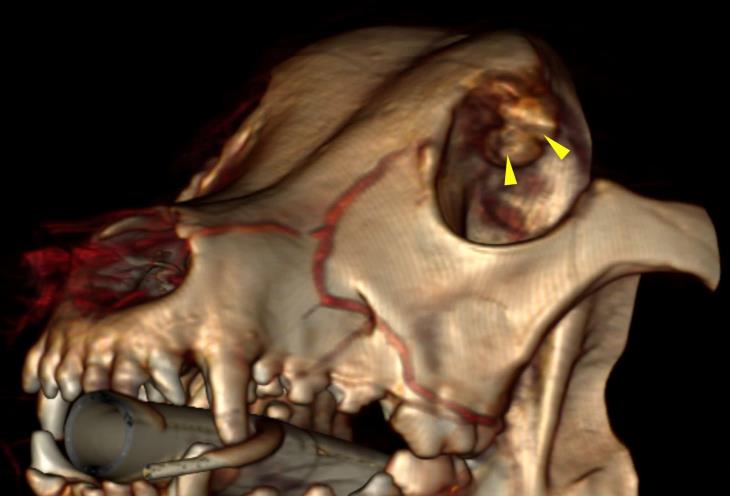
A primary neoplastic disease (osteosarcoma) was diagnosed. The 3D reconstruction shows the base of the mass, at the level of the orbit (yellow arrowheads).
The primary use of CT imaging is of the head of dogs and cats with neurological or nasal diseases. Technologic advances in hardware and software, opened up new possibilities, including abdominal and thoracic imaging, and high definition scanning of lungs and bones.
CT is now widely used in veterinary practice and has become an essential asset to veterinarians in the pursuit of many diagnoses. Long body parts can be scanned within a couple of seconds in amazing detail and accurate contrast resolution.
Thanks to the possibility of reformatting the images in any plane, or as three-dimensional projections, a better representation of the structural anatomic relationships can be achieved.
CT’s diagnostic power is comparable with, or superior to, other imaging modalities such as ultrasound and MRI for many disorders and has immense potential as a rapid and efficient diagnostic tool for a wide range of indications.