Categories
What’s your diagnosis? Computed Tomography Case 4
How Computed Tomography can help us in the diagnosis of diseases of the head
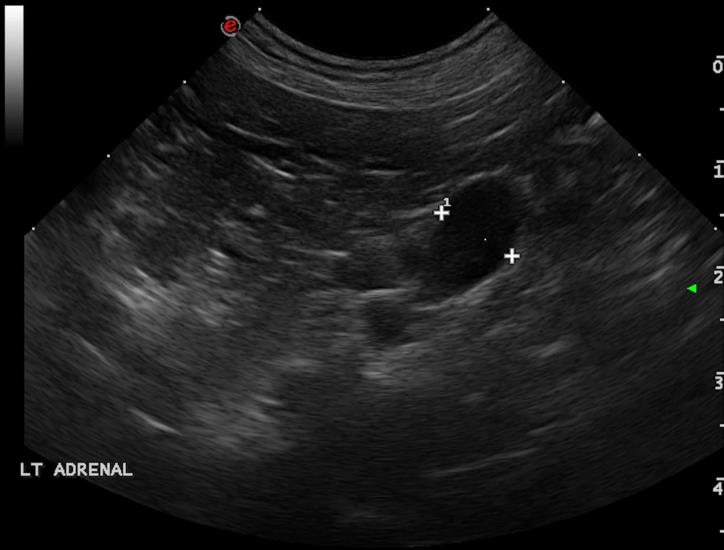
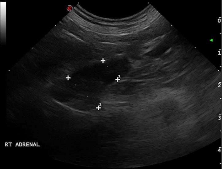
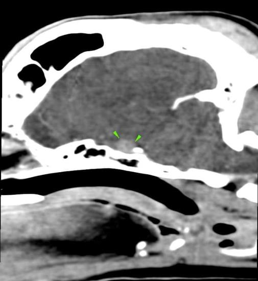
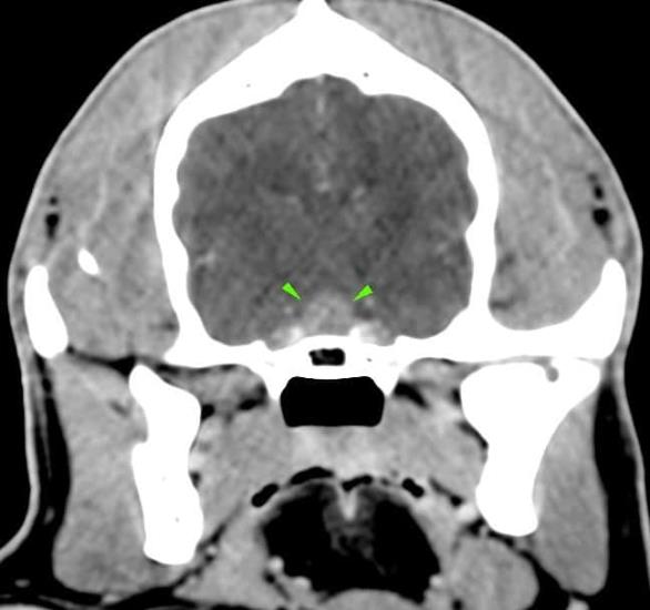
A 8-year-old cocker spaniel was referred for polyuria/polydipsia and polyphagia. The abdominal ultrasound revealed bilateral symmetric adrenal enlargement (images below). A CT scan of the head was performed. A round, 7mm, contrast-enhancing nodule was found at the level of the pavement of the sella turcica, in the hypophyseal fossa (green arrowheads).
Scroll down for the diagnosis…
A final diagnosis of pituitary-dependent hyperadrenocorticism was made.
The primary use of CT imaging is of the head of dogs and cats with neurological or nasal diseases. Technologic advances in hardware and software, opened up new possibilities, including abdominal and thoracic imaging, and high definition scanning of lungs and bones.
CT is now widely used in veterinary practice and has become an essential asset to veterinarians in the pursuit of many diagnoses. Long body parts can be scanned within a couple of seconds in amazing detail and accurate contrast resolution.
Thanks to the possibility of reformatting the images in any plane, or as three-dimensional projections, a better representation of the structural anatomic relationships can be achieved.
CT’s diagnostic power is comparable with, or superior to, other imaging modalities such as ultrasound and MRI for many disorders and has immense potential as a rapid and efficient diagnostic tool for a wide range of indications.