Categories
What’s your diagnosis? Computed Tomography Case 5
How Computed Tomography can help us in the diagnosis of diseases of the head
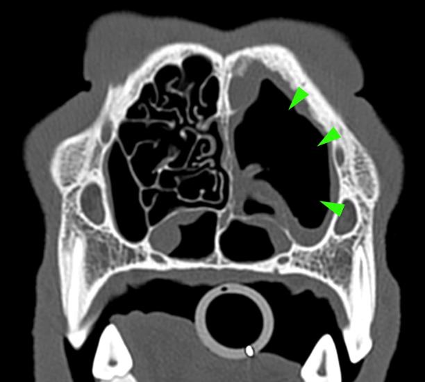
A 3-year-old Siberian husky was presented for chronic nasal discharge. A CT scan of the head showed marked loss of turbinates in the rostral aspect of both nasal cavities, more severe on the left side (green arrowheads).
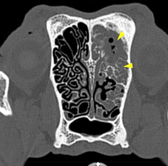
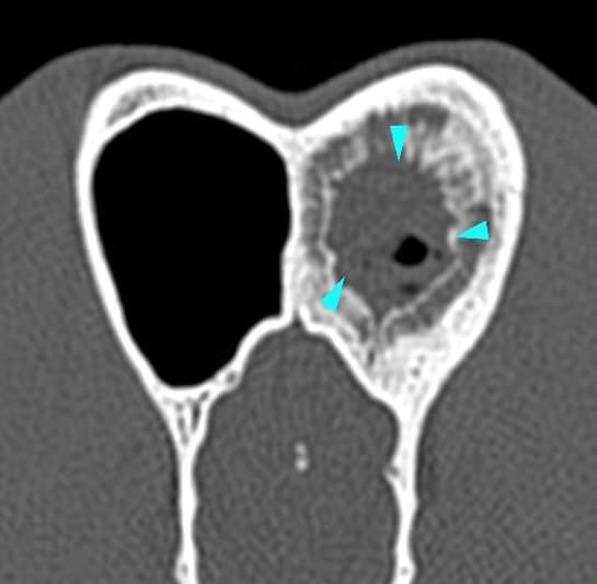
A large amount of fluid was occupying the caudal aspect of the left nasal cavity (yellow arrowheads). There was no lysis of the nasal septum, vomerine groove, ethmoid bone or cribriform plate. The left frontal sinus contained a large amount of fluid-attenuating material, surrounded by a severe ‘lamellar’ periosteal reaction (blue arrowheads).
Scroll down for the diagnosis…
A final diagnosis of fungal rhinitis (aspergillosis) was made.
The primary use of CT imaging is of the head of dogs and cats with neurological or nasal diseases. Technologic advances in hardware and software, opened up new possibilities, including abdominal and thoracic imaging, and high definition scanning of lungs and bones.
CT is now widely used in veterinary practice and has become an essential asset to veterinarians in the pursuit of many diagnoses. Long body parts can be scanned within a couple of seconds in amazing detail and accurate contrast resolution.
Thanks to the possibility of reformatting the images in any plane, or as three-dimensional projections, a better representation of the structural anatomic relationships can be achieved.
CT’s diagnostic power is comparable with, or superior to, other imaging modalities such as ultrasound and MRI for many disorders and has immense potential as a rapid and efficient diagnostic tool for a wide range of indications.