Categories
What’s your diagnosis? Computed Tomography Case 6
How Computed Tomography can help us in the diagnosis of diseases of the head
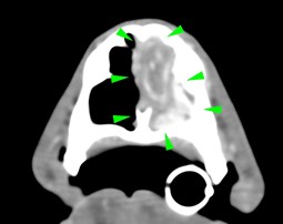
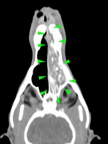
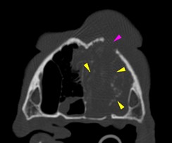
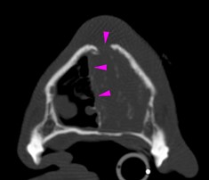
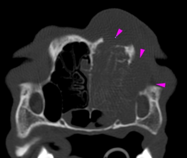
An 11-year-old cocker spaniel was referred to Eastcott Referrals for a chronic nasal discharge and soft tissue swelling in the dorsal aspect of the nose. A CT scan of the head showed a complete obstruction of the left nasal cavity, due to the presence of an amorphous soft tissue mass, extending through the left nasal meatus, left nasal conchae, left sided endoturbinates, left sphenoidal sinus and left frontal sinus (green arrowheads). Bilateral disruption of the turbinates was visible (yellow arrowheads), along with lysis of the left frontomaxillary suture, palatine suture, left rostral portion of the cranium (purple arrowheads) and nasal septum, which was also displaced to the right side
Scroll down for the diagnosis…
A final diagnosis of nasal carcinoma was made
The primary use of CT imaging is of the head of dogs and cats with neurological or nasal diseases. Technologic advances in hardware and software, opened up new possibilities, including abdominal and thoracic imaging, and high definition scanning of lungs and bones.
CT is now widely used in veterinary practice and has become an essential asset to veterinarians in the pursuit of many diagnoses. Long body parts can be scanned within a couple of seconds in amazing detail and accurate contrast resolution.
Thanks to the possibility of reformatting the images in any plane, or as three-dimensional projections, a better representation of the structural anatomic relationships can be achieved.
CT’s diagnostic power is comparable with, or superior to, other imaging modalities such as ultrasound and MRI for many disorders and has immense potential as a rapid and efficient diagnostic tool for a wide range of indications.