Categories
Hock Fracture Repair in Dog with Osteochondritis Dissecans
Repair of hock fracture secondary to osteochondritis dissecans of the medial talar ridge
This hock fracture was secondary to osteochondritis dissecans of the medial talar ridge and was repaired with a lag screw and trans-articular external skeletal fixator.
Case history
An 18-month old male Labrador retriever was presented with a 4/5 right hindlimb lameness after getting up awkwardly and twisting suddenly to the right. Pain and swelling was found on palpation of the right hock joint. A CT scan of the hindlimb revealed an osteochondritis dissecans lesion of the medial talar ridge. However, there was also a minimally displaced fissure fracture extending adjacent to the medial cortex or the medial talar ridge. A caudomedial approach to the medial talar ridge was made. The OCD lesion and associated osteochondral flap was identified and debrided. However, unusual amounts of blood were present within the joint at the time of arthrotomy suggestive of an acute fracture. Close inspection and gentle stress on the joint allowed identification and slight displacement of the fissure fracture running parallel to the medial cortex of the medial talar ridge. A 2mm lag-screw was placed across the fissure fracture from the medial side and recessed into the medial cortex to prevent interference with the medial malleolus. The repair was protected with a trans-articular external skeletal fixator for three weeks. Six weeks after surgery he was using the leg with minimal evidence of lameness and only a mild reduction in the range of motion within the joint. Repeat radiographs showed the screw in position and no sign of instability of the hock.
This is a highly unusual fracture and appears to have occurred as a result of the stress riser created by the presence of an OCD lesion of the medial talar ridge. CT was extremely useful both for the identification of the fracture as well as planning of the precise positioning of the lag screw used to repair it. Application of a TESF is a very useful and adaptable technique which allowed good weight bearing on the limb throughout the recovery period. This was especially important for this dog who has also been treated for bilateral elbow OCD, bilateral medial coronoid disease and bilateral hip dysplasia.
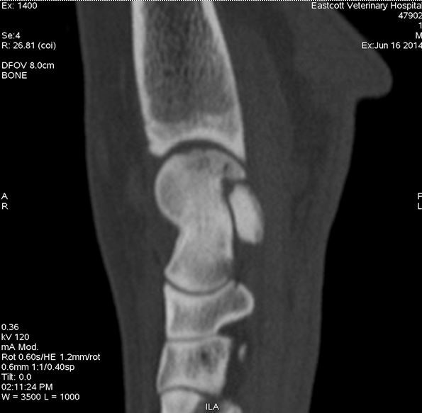
Figure 1
Sagittal image of hock showing OCD lesion of the medial talar ridge
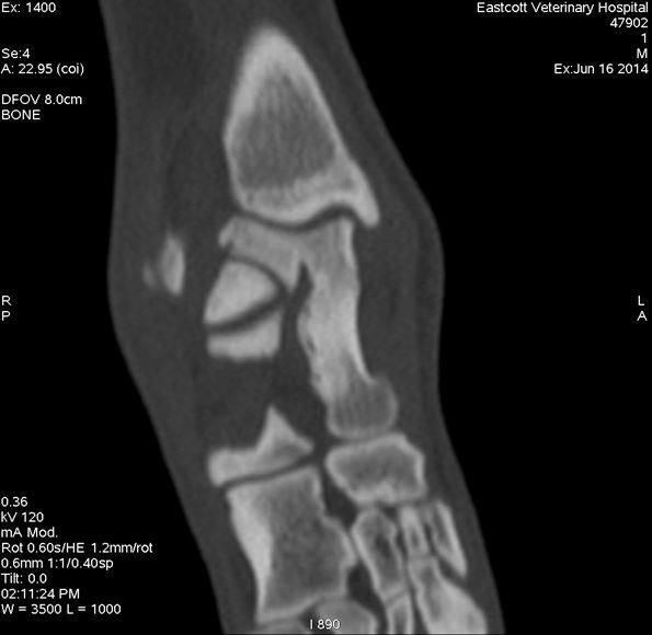
Figure 2
Dorsal plane image through the medial talar ridge showing fissure fracture parallel to medial cortex
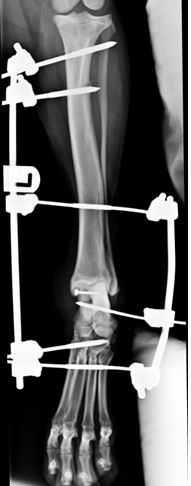
Figure 3
Post-operative radiograph showing lag screw repair being protected by a trans-articular fixator
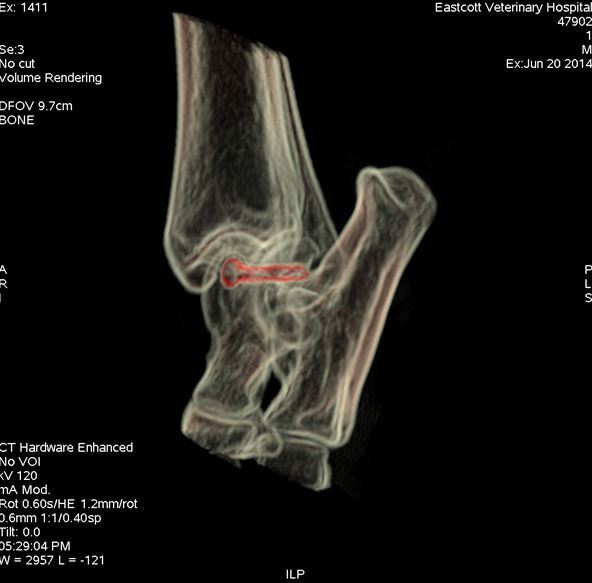
Figure 4
Post-operative CT scan showing position of screw (red) stabilising medial talar ridge fracture

Surgery was carried out by Duncan Barnes MA VetMB CertSAS MRCVS. Duncan is happy to discuss or give advice on all matters relating to small animal veterinary orthopaedics. Please phone 01793 528341 or email Eastcott Referrals