Categories
Pantarsal arthrodesis in a Dog
Pantarsal arthrodesis in a Greyhound dog using a dorsal plate for the treatment of a highly comminuted fracture of the medial talar ridge
A 5-year-old female Greyhound was presented with a non-weight bearing lameness following a fall at high speed.
The leg was markedly swollen with severe bruising and gross instability of the hock joint.
Radiographs revealed a comminuted non-reducible fracture of the medial talar ridge and a displaced fracture of the distal fibula.
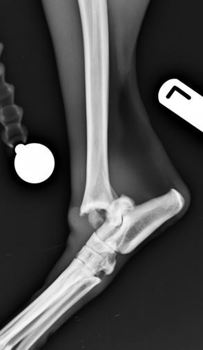
Figure 1. Comminuted talar and distal fibular fractures lateral view
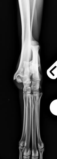
Figure 2. Comminuted talar and distal fibular fractures craniocaudal view
A craniomedial approach to the hock joint was made and upon inspection of the fracture site it was evident that it would not be possible to reconstruct and repair the talar fracture. A decision was therefore made to perform a pantarsal arthrodesis using a specifically designed dorsal pantarsal arthrodesis plate. The plate features 3.5mm screws in the proximal part of the plate and 2.7mm screws distally in the metatarsal. This provides an extremely rigid stable fixation. Cartilage was removed using a high speed burr from the articular surfaces. The fractured parts of the talus were morselised, mixed with further cancellous bone graft taken from the proximal tibia and demineralised bone matrix and used to fill the joints spaces. The plate was applied and the skin closed routinely.
She made a good recovery from the anaesthetic and no further problems were seen during recuperation. The excellent biomechanical stability of the repair meant that a cast was not needed post-operatively, avoiding cast associated complications.
Six-weeks following surgery radiographs revealed excellent progression of arthrodesis and no evidence of implant associated problems. Normal exercise was gradually re-introduced at this point. Pantarsal arthrodesis was successful for salvaging a pain-free hindlimb after a potentially catastrophic fracture of the articular surface of the talus of a greyhound.
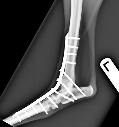
Figure 3. Six-weeks post-operative lateral view
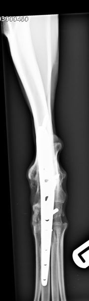
Figure 4. Six-week post-operative craniocaudal view

Surgery was carried out by Duncan Barnes MA VetMB CertSAS MRCVS. Duncan is happy to discuss or give advice on all matters relating to small animal veterinary orthopaedics. Please phone 01793 528341 or email Eastcott Referrals