Categories
Shoulder Arthrodesis Using CT Generated 3D Bone Model
Shoulder arthrodesis in a miniature poodle using a CT generated 3D bone model
A 9-year-old miniature poodle was referred for the treatment of recurrent luxation of the right shoulder. Examination under general anaesthetic revealed a markedly increased abduction angle and gross instability of the right shoulder joint. Due to chronic non-weight bearing lameness there was very poor muscle around the shoulder joint. After two failed attempts had been made to stabilise the joint using sutures, the decision was made to perform a shoulder arthrodesis. This procedure can be an excellent way to resolve chronically painful shoulder conditions whilst maintaining a good level of forelimb function.
A CT scan of the humerus and scapula was performed and from this a three-dimensional life size model was produced using 3D printing technology (3D model arranged through Vet CT Specialists).
This allowed rehearsal of the surgery and precise contouring of the plate used prior to surgery. The benefits of this approach include greater accuracy of plate contouring, markedly reduced surgical time and more predictable post-operative limb alignment.
During surgery the articular cartilage was debrided from the joint using an air-bur and the joint was temporarily stabilised using an arthrodesis wire. A trans-articular lag screw was applied after packing the joint with cancellous bone graft. The pre-contoured 2.0mm locking SOP plate (Orthomed) was then applied across the joint.
Within a few weeks of surgery the dog was bearing weight well on the operated leg and was comfortable enough to stop receiving NSAIDs.
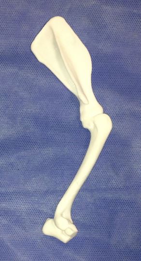
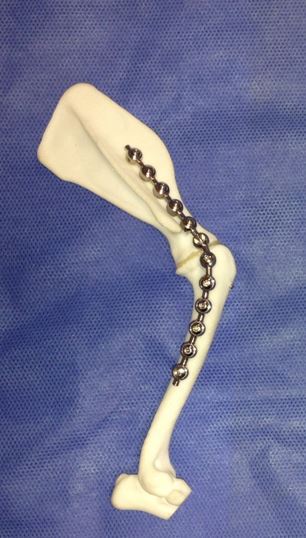
Fig 1: Bone model created from the CT scan Fig 2. A second model used to rehearse the surgery and contour the plate appropriately
Radiographs taken 8 weeks after surgery showed good progression of arthrodesis and no evidence of any implant associated problems.
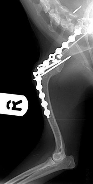
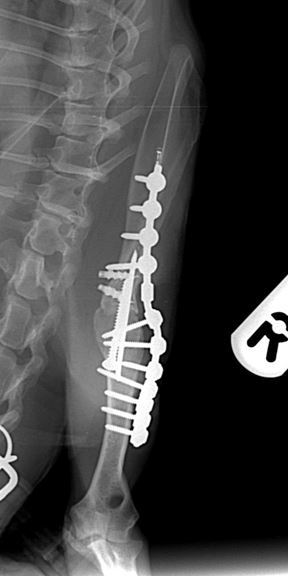
Figures 3 & 4. Mediolateral and caudocranial radiographs of the arthrodesed shoulder 8 weeks after surgery, showing good progression of arthrodesis and good alignment of the shoulder and elbow joints with the shoulder at a standing angle.
Surgery was carried out by Duncan Barnes . Duncan is happy to discuss or give advice on all matters relating to small animal veterinary orthopaedics. Please phone 01793 528341 or email Eastcott Referrals
3D life size was arranged though Vet CT Specialists
Examples of orthopaedic cases seen by Duncan at Eastcott Referrals