Categories
Why we use TPLO for treatment of cranial cruciate ligament disease

Cruciate ligament disease is the most common orthopaedic problem encountered in dogs. The cranial cruciate ligament’s primary role is to control cranial movement of the tibia relative to the femur, as well as controlling rotation about the stifle joint. Cranial cruciate ligament disease is most commonly a result of degenerative changes within the ligament. Dogs may present with a complete rupture of the ligament or with stifle pain associated with a partially torn cruciate ligament. We would recommend surgical treatment in the majority of dogs and cats to get the best function possible. However, where surgical treatment is not possible, conservative treatment remains a reasonable option.
A variety of surgical techniques have been developed to treat cranial cruciate ligament disease. At Eastcott Referrals each patient is assessed to determine the most appropriate treatment for the individual. Most dogs will be treated with a tibial plateau levelling osteotomy (TPLO), whilst other dogs are better suited to a cranial closing wedge ostectomy. For some dogs an extracapsular suture technique may be the best option.
Tibial Plateau Levelling Osteotomy (TPLO)
TPLO has been shown to be a robust and reliable procedure for dogs of all sizes. Most dogs are weight bearing well on the operated leg the day following surgery and benefit from the early limb use this allows. The procedure is designed to change the biomechanics of the stifle joint such that it neutralises cranial tibial thrust during the stance phase effectively doing away with the need for a cranial cruciate ligament. TPLO is suitable for dogs at every stage of cruciate disease from early partial tears, acute traumatic cruciate tears to chronic arthritic stifles. We expect 95% of dogs to have a good or excellent outcome following TPLO with a low rate of complications. A recent study by Krotscheck 2016 compared extracapsular suture, tibial tuberosity advancement and TPLO, using force plate assessment at 8 weeks, 6 months and 12 months. Whilst dogs having TTA had slightly better weight bearing at 8 weeks, TPLO was the only surgery able to return dogs to normal weight bearing at 6 and 12 months after surgery at both a walk and a trot. With this evidence we consider TPLO to be the best option for the treatment of cruciate disease in dogs.
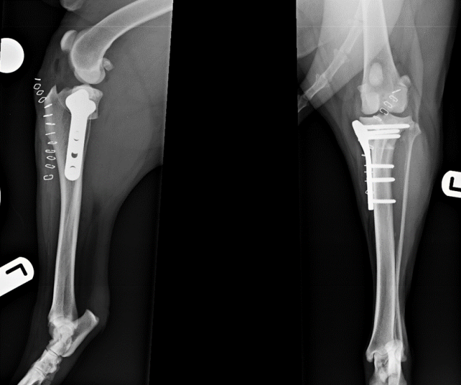
Figure 1. Tibial plateau levelling osteotomy using a broad locking plate and screws in a 45kg Labrador
Cranial closing wedge ostectomy (CCWO)
CCWO achieves tibial plateau levelling in a different way to TPLO but will have similar biomechanical effects. We use CCWO for dogs with proximal tibial deformity leading to angulation between the proximal and distal tibial axes. This includes many small breed dogs with cranial cruciate ligament disease. It is an effective way to treat cranial cruciate ligament disease in juvenile dogs where implants cannot be placed across the open physes, allowing unimpeded continued growth of the tibia (Figure 2).
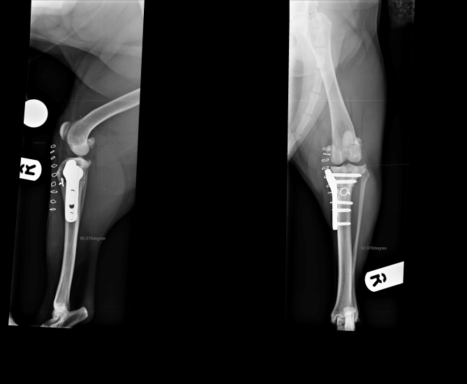
Figure 2. Treatment of cranial cruciate ligament rupture in a West Highland white terrier using a cranial closing wedge ostectomy stabilised with a locking plate and screws
Complex cases
Some cases of cranial cruciate ligament disease are more complicated than others including:
- Juvenile animals (Figure 3)
- Combined cranial and caudal cruciate ligament rupture
- Concomitant medial patellar luxation (Figure 4)
- Concomitant angular limb deformity
- Extreme tibial plateau angle (Figure 5)
- Tibial varus or valgus
All of these can be treated effectively by adapting our standard techniques. For the cases with angular limb deformity including extreme tibial plateau angle (>34 degrees), CT of the hindlimbs allows us to perform an accurate assessment of all aspects of hindlimb alignment. This facilitates planning of the most appropriate surgery, reducing surgical time and leading to excellent clinical results.
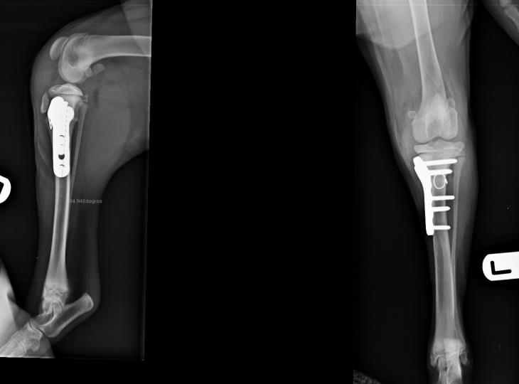
Figure 3. Treatment of cranial cruciate ligament avulsion in a 4-month-old Golden retriever using a cranial closing wedge ostectomy
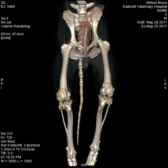
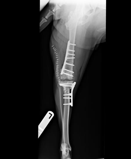
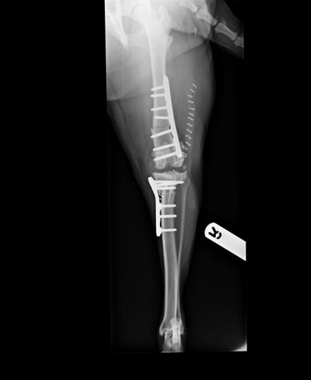
Figure 4. Staged bilateral surgical correction of combined distal femoral varus, grade ¾ medial patellar luxation and cranial cruciate ligament rupture using distal femoral ostectomy, block recession trochleoplasty and transposed tibial plateau levelling osteotomy
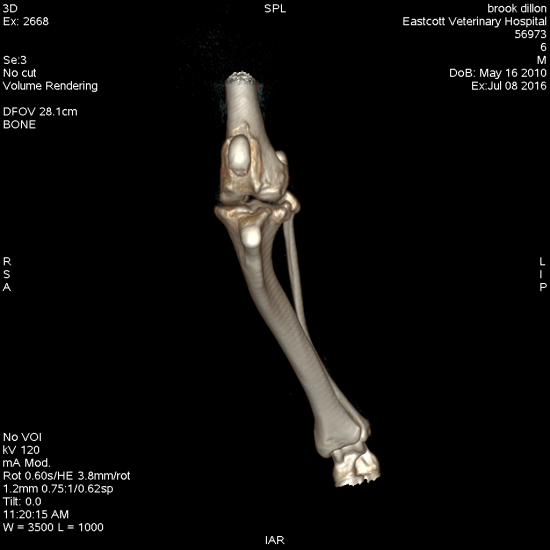
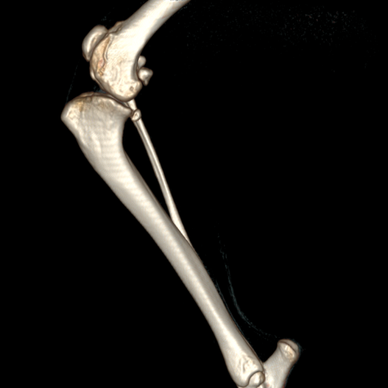
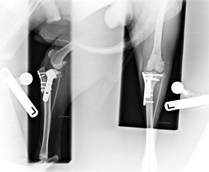
Figure 5. Treatment of combined excessive tibial plateal angle (56 degrees) and severe tibial valgus deformity using a combined TPLO and cranial and medial closing wedge ostectomies
Duncan and the orthopaedic referral team are happy to take your advice calls and emails