Categories
Aggressive Keratomalacia in a White Boxer Dog
Keratomalacia in Small Animals
Keratomalacia, or “corneal melting”, is a process of degeneration and liquefaction of the corneal stroma, which is mainly associated with bacterial infection, typically Streptococci and Pseudomonas species, and the neutrophilic response to the infection. Some bacteria as well as the responding neutrophils have the ability to secrete enzymes such as proteases, collagenases and elastases, which attack structural collagen and glycosaminoglycans of the cornea. Additionally they can break down the intrinsic inhibitors of these enzymes within the cornea, hence having the ability to suddenly massively accelerate the process of damage to the cornea and many cases deteriorate rapidly and present as ophthalmic emergencies. Some cases are responsive to intensive medical management, if recognised and presented early enough, but a percentage of cases are so extensive or deep that they present an immediate threat to corneal integrity and require surgery.
Brachycephalic breeds of dogs are pre-disposed to keratomalacia, but the condition can be seen in all breeds of cats and dogs.
Keratomalacia Surgical Options in Small Animals
Surgical options to help save eyes suffering from severe keratomalacia should aim to introduce blood vessels, as well as provide tectonic strength and options include an initial keratectomy to remove as much of the damaged tissue as possible together with a conjunctival pedicle graft, corneo-conjunctival transposition graft and a 360-degree conjunctival graft.
Prognosis
These cases carry a high risk of rupture and should be given a guarded prognosis, but the outcome can still be surprisingly good if surgery is done in a timely fashion.
What Happens if the Whole Cornea is Malacic?
Cases with larger areas of deep keratomalacia may not be suitable for grafting due to insufficient available cornea and conjunctiva, but can respond well to a 360-degree conjunctival graft and should be given this option in addition to for instance enucleation.
Below is an example of an extensive and complicated melt, which required urgent surgical attention:
Diffuse, progressive Keratomalacia in a 5 year old white Boxer dog
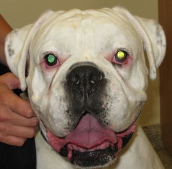
Clinical Symptoms
Jake presented with a painful left eye with the following clinical symptoms:
- Marked epiphora
- Blepharospasm
- Marked episcleral and conjunctival hyperaemia
- 360 degree ciliary flush
- Central stromal abscess/WBC infiltrate
- Deep corneal melting ulcer (note the circular shape and deep edges)
- Marked corneal oedema
- Poor vision due to the corneal clouding
- Severe reflex uveitis is suspected, including miosis
Diagnostic Tests
- Schirmer tear test – high normal fellow eye and visibly high affected eye
- Cytology and culture not done due to fragility of the cornea and ‘sealed’ stromal abces, but should be done as far as possible in all cases as multi-resistad cases are not uncommon and should screen for fungal hyphae
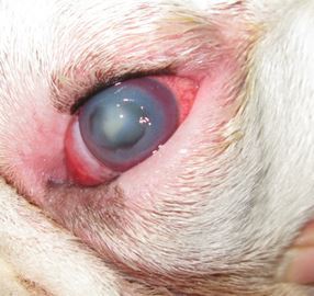
Advanced keratomalacia of the left eye
Surgery
A keratectomy was performed under the operating microscope to remove as much malacic tissue as possible and provide fresh edges for a large ventral corneoconjunctival transposition (CCT) graft, bringing bloodvessels and tectonic strength into the central cornea. 9/0 PGA was used for the sutures.
Medication
- Exocin e4h
- Atropine SID x 3 days
- Ronaxan SID
- Metacam SID
3 Days Post Surgery
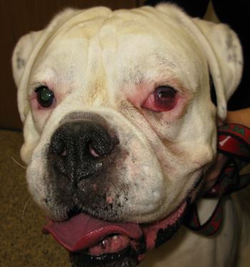
3 days post surgery
The eye was comfortable with only mild epiphora and the graft settling. The conjunctival blood vessels looked healthy. There was some bruising on the corneal section due to disruption of the corneal vessels (the thickness was reduced surgically). The horizontal white line is the transposed limbus, which remains visible long-term following this type of graft.
7 Weeks Post Grafting
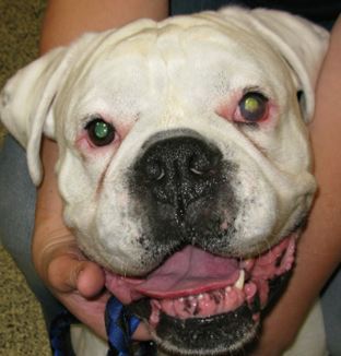
The 9/0 sutures resorbed around 6 weeks post-operatively.
The eye has useful vision through the graft and the sutures have resorbed. The eye was put on steroid eye drops to help improve vision further and reduce scarring.
11 Weeks Post Surgery
At the final check up, the corneal clarity had improved significantly and all drugs were stopped. Jake has regained good, functional vision in his left eye.
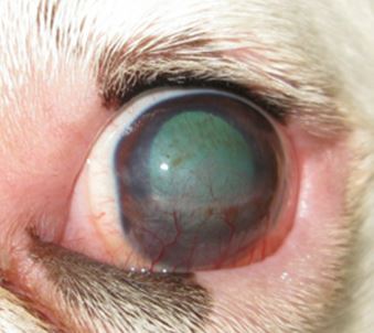
11 weeks post operatively, A good surgical outcome, considering the severity of damage to this cornea

If you think you have a suitable case that you would like to refer, or if you would like any more information, please contact Ida Gilbert BVSc CertVOphthal MRCVS on 01793 528341 or e-mail: Eastcott Veterinary Referrals
Other Ophthalmology Cases Commonly Seen
Eastcott Veterinary Referrals CPD Courses
How to Refer a Case to Eastcott Veterinary Referrals