Categories
Avoiding Ruptured Diabetic Cataracts in Dogs
Ruptured diabetic cataracts: An avoidable emergency
A recent review of our cataract surgery patients found that approximately 50% of cases were diabetic dogs. Functional vision was achieved in over 95% of these cases, however some of our diabetic patients were not suitable for cataract surgery due to potentially avoidable lens rupture and retinal detachment. Vision can be saved in some cases of lens capsule rupture if surgery can be performed promptly.
Diabetic Cataracts in Dogs – The enemy: Aldose reductase
High levels of glucose in the blood overwhelm the normal metabolic pathway in the lens leading to the formation of sorbitol. Sorbitol cannot escape the lens so builds up inside it and draws water in from outside of the lens capsule. This process is facilitated by the enzyme aldose reductase (AR). Levels of AR vary from species to species (cats have very little so are unlikely to suffer from diabetic cataracts) and it is thought that the amount of AR present in the lens varies from individual dog to dog. It appears that there are two distinct groups of diabetic cataract patients: Group 1. Dogs that form cataracts in the first few months following diagnosis and in which the cataracts form very quickly and Group 2. Dogs with a later and more gradual cataract formation.
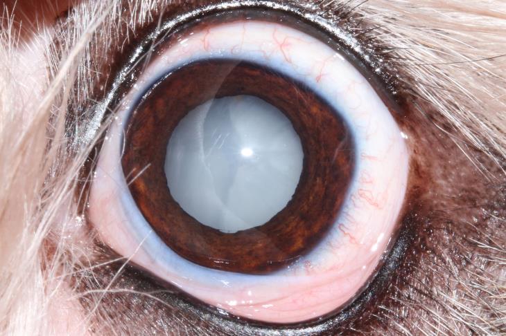
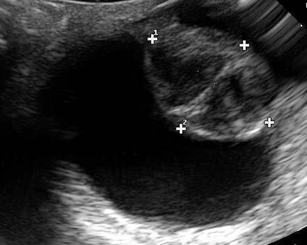
Fig 1 and 2: Mature diabetic cataract showing a swollen lens and clear water clefts forming and an ultrasonograph of a similar lens.
Diabetic Cataracts in Dogs – The problem: Lens capsule rupture
Group 1 cases are thought to have higher levels of AR in their lenses and can become genuine emergency
cases. The lens can swell so dramatically that the lens capsule ruptures which in turn leads to a severe uveitis and risks the blinding secondary consequences of retinal detachment and eventual glaucoma.
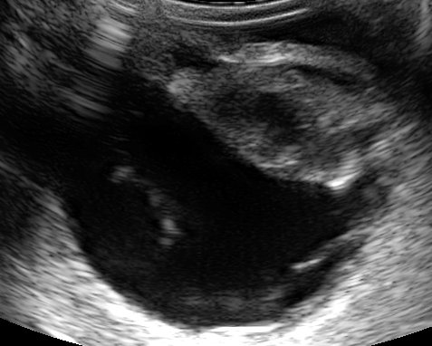
Figure 3: This ruptured lens has an irregular shape with a flattened posterior outline. Retinal detachment is also present.
Diabetic Cataracts in Dogs – The solution: Prompt referral
Currently the only effective way to deal with these cases is to remove the source of the inflammation: i.e. the cataractous lens. This is often a race against time and the urge to completely stabilise your diabetic patient prior to surgery. If lens capsule rupture has occurred they need to have their lenses removed urgently to avoid permanent blindness. If your diabetic patient suddenly develops mature cataracts and your owner is in a position to consider cataract surgery a prompt assessment by a veterinary ophthalmologist could save their vision. Treatment with NSAIDs (systemic and topical) prior to
surgery is desirable (2 to 7 days of treatment is ideal). As soon as any DM cataract is noted it is advisable to begin anti-inflammatory treatment (e.g. Ketorolac OU BID and Meloxicam PO SID).
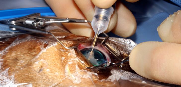
Figure 4: Cataract being aspirated up the phacoemulsification probe

Diabetic Cataracts in Dogs – The future: Kinostat?
Kinostat is an aldose reductase inhibitor. It is currently being licensed in the USA as an eye drop for dogs that has been shown to reduce cataract formation in diabetic patients. It is not currently available in the UK.
by Mark Ames BVSc CertVOphthal CertVDI MRCVS. Mark is happy to discuss all matters relating to Small Animal Ophthalmology.