Categories
Bilateral Pyogranulomatous Meibomianitis In a Dog
Bilateral Pyogranulomatous Meibomianitis in a Dog
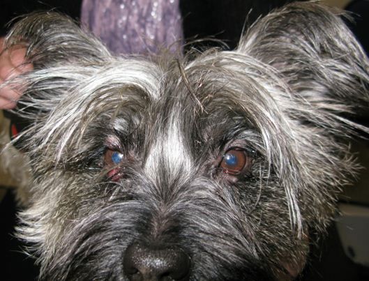
A 2.5-year-old Cairn Terrier was referred for excision of two enlarging eyelid masses in the right eye. They had been present for about a month. On presentation, the left eyelids had also started to develop nodular swellings and there was a mucoid to mucopurulent discharge present.
Anterior view of both eyes a week after instigating treatment.
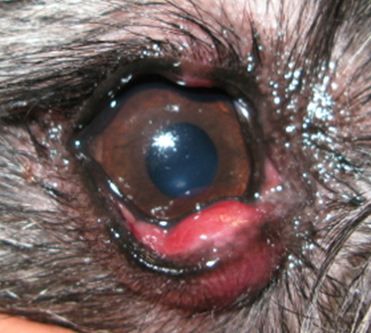
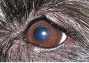
Close up of the right eye.
Close up of the right eye.
Active meibomianitis with pyogranulomas, associated with Meibomian gland rupture, was identified and initially medical treatment was instigated together with hot compresses.
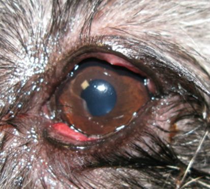
The larger granulomas were developing marked fibrosis and surgery was required to de-bulk and curettage these. Full thickness excision was avoided.
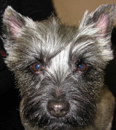
4 days after surgery to debulk the swellings. Both eyes are comfortable and there is no discharge. The right lower eyelid will re-pigment in time.
Close up of the left eye, which achieved the best result, as this had a shorter period of active inflammation and scarring. Both eyes are comfortable and a good cosmetic result was achieved.
All of our Ophthalmology cases are seen by Ida Gilbert BVSc CertVOphthal MRCVS. Ida is happy to discuss or give advice on all matters relating to small animal ophthalmology. She can be contacted via email at Eastcott Referrals