Categories
Cholesteatoma in Dogs
Cholesteatoma- a New Disease?
Cholesteatoma is an epidermoid cyst that forms within the middle ear cavity. Contrary to what the name suggests, it is non-neoplastic and has nothing to do with cholesterol or fat. Rather, it is charaterised by a cornifying stratified squamous epithelium which produces keratin leading to a slowly expansile lesion within the tympanic bulla. The aetiology is incompletely understood but, although congenital forms are possible, they are most likely a result of chronic otitis externa in which keratinizing stratified epithelium has had the opportunity to seed and establish within the middle ear chamber.
Although in theory the cyst is benign, it can cause a range of signs relating to its position and size. Symptoms therefore include signs of otitis media – head tilt, nystagmus, circling etc but animals can also present as dogs who are reluctant to fully open their mouths and/or are reluctant to eat and this is due to encroachment of the cholesteatoma on the temporomandibular joint (TMJ). We have also seen some dogs which presented with dysphagia due to orpharyngeal compression from ventral outgrowth from the bulla. The lesions can also extend medially and erosion of the cranial vault can lead to otitis interna and even meningoencephalitis.
The treatment of choice is surgical removal of the keratin contents together with the secretory lining responsible for the lesion and this involves either total ear canal ablation and lateral bulla osteotomy or ventral bulla osteotomy (or both!). Surgery can be challenging as the distorted anatomy makes it hard to remove all of the responsible epithelium and this leads to recurrence which is reported in approximately 50% of cases (Hardie et al 2008). Recurrence is most likely in dogs with either neurological signs, bone lysis or inability to open the mouth.
With the increasing availability of advanced imaging (CT, MRI etc) increasing numbers of Cholesteatoma are being diagnosed and so it is being regarded as an emerging disease. There is no reason to think that this is the case, however with cholesteatoma recognised in 10% of cases of otitis media in the early 1990’s (Little et al 1991). We are now better at diagnosing the condition, however, and early detection of these lesions leads to better clinical outcomes. We therefore advise considering CT examination of dogs with persistent otitis externa and early surgical intervention in dogs which are diagnosed with this condition.
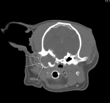
Fig 1:
Transverse CT showing expanded and irregular, tissue filled left tympanic bulla
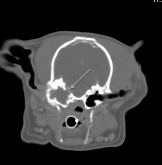
Fig 2:
Transverse CT of the same dog in Fig1 showing erosion into the cranial vault and meningeal enhancement (meningitis)
By Tim Charlesworth

If you think you have any small animal soft tissue case that you would like to refer or discuss, please contact Tim on 01793 528341 or e-mail: Eastcott Veterinary Referrals