Categories
Dog With Bilateral Diabetic Cataracts and (unrelated) Benign Limbal Melanoma
Bilateral Diabetic Cataracts followed by (unrelated) Benign Limbal Melanoma in an 8-year old dog
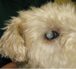
This 8 year old Fox Terrier has bilateral cataracts
Close up of right eye pre-cataract surgery
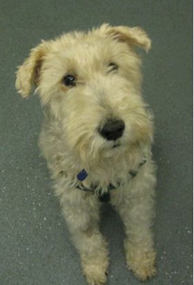
Dog 1.5 week after bilateral cataract surgery
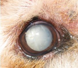
Close up of right eye 7 weeks after cataract surgery
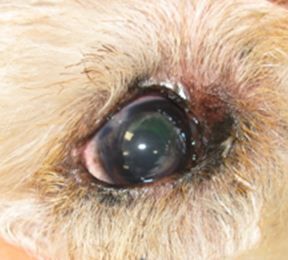
6 Months Post Cataract Surgery Examination
Both of this dog’s eyes did very well, but at the 6-months check post phacoemulsification, the right eye was causing minor bother to the dog and a pigmented swelling had developed ventromedially below the Nictitans membrane. A presumed diagnosis of benign limbal melanoma was made and we decided to proceed to surgery. A lamellar excisional biopsy was done, followed by a double freeze-thaw cycle of cryosurgery according to the protocol described by Featherstone et al Vet Ophthal 2009 Nov-Dec; 12 Suppl 1: 65-72 (Efficacy of lamellar resection, cryotherapy and adjunctive grafting for the treatment of canine limbal melanoma).
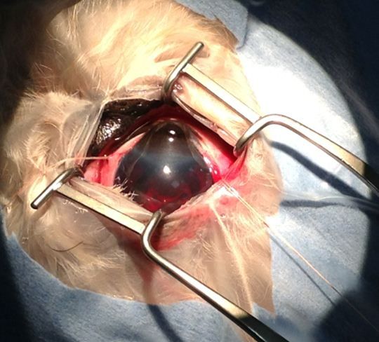
The limbal melanoma can be seen extending into the cornea and causing gross thickening and distortion of the normal corneal curvature.
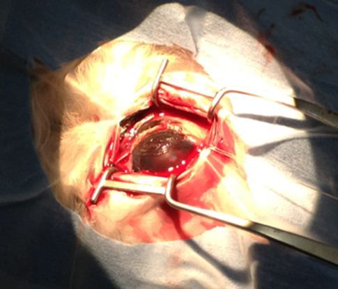
Right eye immediately after excision, but before cryosurgery and grafting
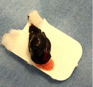
The bulk of the melanoma. Histopathology showed that this was a benign melanoma
2 weeks post excision of the limbal melanoma
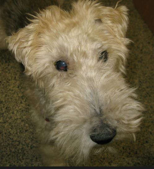
2 weeks post excision of the limbal melanoma in the right eye. The edge of the pink conjunctival graft can be seen ventromedially. Jazz continues to do well 5 months later.
Eastcott Veterinary Referrals offer a full Small Animal Cataract Service. All of our ophthalmology cases are seen by Ida Gilbert BVSc CertVOphthal MRCVS. Ida is is very happy to give advice over the phone or by e-mail (photographs are very helpful if you want to discuss a specific case).