Categories
Portosystemic Shunts in Small Animals
Portosystemic Shunts in Small Animals at Eastcott Referrals
About Portosystemic Shunts in Dogs & Cats
Portosystemic Shunts (PSS) are congenital, abnormal communications between the portal and systemic circulations. This allows toxins, which would usually be removed by the liver, to enter the general (systemic) circulation and exert a number of adverse effects. Numerous body systems are affected causing a wide range of symptoms to develop. Symptoms can be neurological (e.g. lethargy, depression, episodic blindness, head pressing, seizures), gastrointestinal (e.g. vomiting, diarrhea, ascites), urinary (e.g. dysuria, polyuria, polydipsia) or affecting general well being (e.g. poor growth rate, prolonged effect of drugs administered).
PSS’s can be seen in both cats and dogs. Certain breeds are at increased risk of PSS including Himalayan cats, Maltese, Cairn, Yorkshire, Silky, Jack Russell and West Highland White Terriers, Miniature Schnauzers, Irish Wolfhounds, Golden Retrievers, Old English Sheep Dog and Australian Cattle dogs. Affected dogs and cats should not be used for breeding.
The shunt location can either be intrahepatic or more commonly extrahepatic.
Treatment of Portosystemic Shunts in Dogs & Cats
Treatment is either symptomatic/medical (e.g. low protein diet, antibiotics and lactulose) or surgical with longer survival times reported after surgical management.
Diagnosis of Portosystemic Shunts
Diagnosis is usually made on the basis of a combination of suggestive serum biochemistry and haematological findings and compatible clinical signs. The diagnosis is confirmed using imaging (usually ultrasound or CT) to identify the abnormal blood vessel. Ultrasound can provide a rapid way to confirm the diagnosis and to visualise the shunt and it can be performed in the conscious patient. CT angiography can also be performed (Fig 1) which is even more sensitive than ultrasound, gives additional information useful to surgical planning but does require a general anaesthetic.
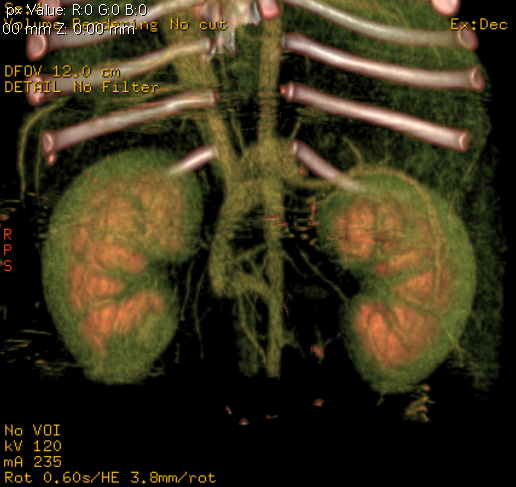
Fig 1: Angiogram of the cranial abdomen showing position of a portocaval shunt
Portosystemic Shunt Surgery in Small Animals
Surgery is performed after a period (typically several weeks) of medical management.
Intraoperative fluoroscopy is used to confirm the presence of a shunt

Fig 2: PSS surgery is performed using intraoperative fluoroscopy
The shunt is identified (Fig 3) and a test ligation performed to confirm identification of the correct vessel and to assess whether or not the patient can tolerate complete ligation of the shunt (Figs 4-5)
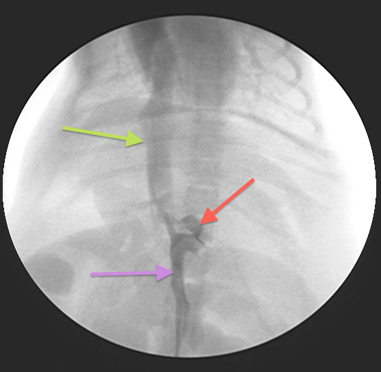
Fig 3:
Intraoperative fluoroscopy (portovenogram) showing the same PSS as in Fig 1: Purple arrow = Portal vein; Green arrow = CVC; Red arrow = PSS
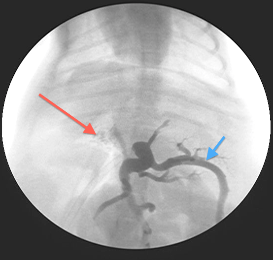
Fig 4:
Portovenogram following test ligation (temporary complete occlusion of the PSS) – most contrast has backed up through the splenic vein (blue arrow) with only some contrast going through the right portal vein into the liver (red arrow). This patient would not tolerate complete ligation of the shunt
.JPG)
Fig 5: Intraoperative appearance of an extrahepatic PSS within the epiploic foramen with a vascular snare placed to perform the test ligation portovenogram
Most animals cannot tolerate complete ligation at the time of surgery as the portal circulation is usually relatively under-developed and unable to cope with the increased blood flow. This would lead to portal hypertension which can be rapidly fatal.
PSS’s in animals unable to tolerate complete occlusion are therefore gradually attenuated by placement of either an Ameroid Constrictor or Cellophane Band around the shunt allowing gradual reduction of the shunt and simultaneous development of the portal vasculature in response to the increased blood flow.
Prognosis
The majority of dogs who undergo surgery go on to do very well although there are significant potential complications all of which are discussed in detail with the owner at the initial consultation. Medical management is gradually discontinued over the 6-8 weeks following the surgery and the dogs can then start to be fed higher-protein diets. Follow up blood samples and ultrasound scans can be used to monitor resolution of the PSS.
Portosystemic Shunt Referrals in Small Animals
At Eastcott we are happy to offer a range of services to help with any portosystemic shunts ranging from ultrasound or CT only appointments through to full case management i.e from imaging through to surgery and postoperative care.
Please do feel free to contact us if you would like any advice about any potential cases that you may have, have any questions or if you would like to make a referral on :01793 528341 or click HERE to email us
By Tim Charlesworth
If you think you have any small animal soft tissue case that you would like to refer or discuss, please contact Tim on 01793 528341 or e-mail: Eastcott Veterinary Referrals