Categories
Subcutaneous Ureteric Bypass or SUB placement in Cats
By Tim Charlesworth
 Ureteric obstruction is an increasingly diagnosed condition in cats with calcium oxalate urolithiasis being the most common cause. Cats often present with vague clinical signs and so the diagnosis can be challenging. Cats often have obstruction of one ureter without showing any significant clinical signs and then present in an advanced stage of disease when the other ureter becomes obstructed. The condition is often misdiagnosed as acute on chronic renal failure (CRF) as cats may have a sudden increase in their azotaemia and it is impossible to differentiate these two conditions without an abdominal ultrasound.
All cases of ureteric obstruction benefit from a period of medical management although this is only successful at fully resolving the ureteric obstruction in a minority of cases. Cats that present with significant hyperkalaemia are usually best treated surgically after an initial 24-48 hours of stabilisation.
Traditional surgical options include ureterotomy (removing the stone directly from the ureter) or flushing it back into the renal pelvis and then performing a pyelotomy/nephrotomy. These techniques sadly carry significant risks of urine leakage, scarring and subsequent stenosis (ureterotomy) or permanent loss of renal function (nephrotomy) and so are no longer recommended for this condition. It is occasionally possible to resect the obstructed ureter if the stone is relatively distal and then re implant the ureter into the base of the bladder but this is again associated with risks of complications (<30%) relating to the ureterovesicular anastomosis.
The relatively high incidence of uroabdomen and stricture formation following ureterotomy and ureterectomy has prompted development of new surgical techniques. Ureteric stents are polyurethane tubes that contain multiple fenestrations. There is a pigtail at either end; one end sits in the renal pelvis and the other in the ureteric opening at the bladder trigone. These stents provide passive ureteric dilation and urine can flow either through them or around them. Stents can, however, be very challenging to place and surgery times can be prolonged. In addition, there is a high rate of dysuria because of the position of the pigtail in the trigone. Other complications include stent fracture, migration and encrustation leading to blockage.
The most recent innovation is the Subcutaneous Ureteric Bypass system (“SUB”). This is an extra-anatomic device consisting of a pigtail nephrostomy tube and a cystostomy tube which are connected via a subcutaneous access port. The SUB therefore allows decompression of the obstructed renal pelvis without addressing the ureteric obstruction itself and effectively acts as a “false ureter”. They can be placed relatively quickly which minimises anaesthetic time for these debilitated patients. Placement does however require intraoperative fluoroscopy and familiarity with interventional radiology techniques. The access port allows postoperative flushing of the system to prevent blockage/ensure long term patency and also allow urine sampling to monitor for urinary tract infections.
SUBs are currently our favoured technique to treat obstructive ureterolithiasis. There is no long term follow up for these cats, however, as the implants simply have not been available for that long. We have placed several in cats over the last couple of years and short term results have so far been very promising. There is sadly no ideal surgical option for treating these cats but the current evidence base suggests that SUBs are the best solution to this increasingly common and potentially life threatening condition in cats.
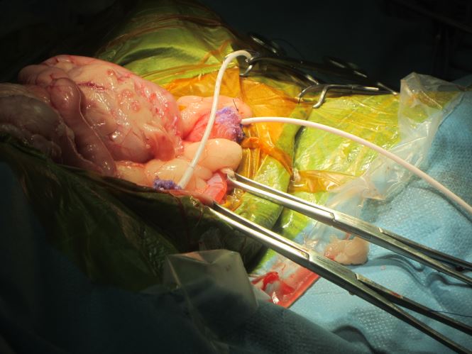
Fig 1: A close up view of the SUB cystostomy and neprhrostomy tubes during placement
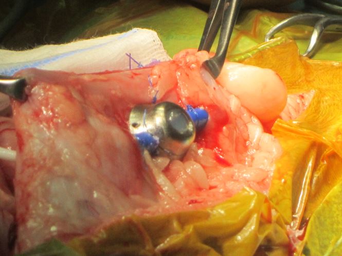
Fig 2: The access port for the SUB. This remains palpable and so can be readily accessed to permit future sampling and flushing of the SUB system
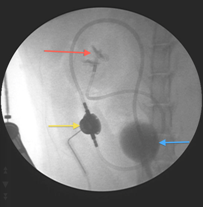
Fig 3: An intraoperative fluoroscopy picture confirming correct placement of the implants. Red arrow = pigtail nephrostomy tube within renal pelvis; Yellow arrow = access port; Blue arrow = bladder (containing contrast)
If you think you have any small animal soft tissue case that you would like to refer or discuss, please contact Tim on 01793 528341 or e-mail: Eastcott Veterinary Referrals