Categories
Untreated Malocclusions in Dogs
Abnormal occlusion is relatively common in dogs and can be associated with the abnormal position of a single tooth or more complex situations associated with discrepancies in the relative lengths of the maxilla and mandibles.
Malocclusions in Dogs
Malocclusions which result in abnormal tooth to tooth or tooth to soft tissue contact require treatment in order to avoid endodontic disease and/or chronic trauma to oral soft tissues.
Case History
In this case a four year old Dogue de Bordeaux presented with a history of crepitus in it’s temporomandibular joints and restricted jaw opening (distance between incisal edges of central maxillary and mandibular incisors of 4.5cm). There was no history of trauma. Clinical examination demonstrated a Class II (mandible shorter than maxilla) malocclusion and abnormal wear patterns on the distal aspect of the maxillary canine teeth and the mesial aspect of the mandibular canine associated with abnormal tooth to tooth contact.
A CT scan showed bilateral temporomandibular joint (TMJ) degenerative disease (right worse than left); right retroarticular process fragmentation; bony proliferation affecting the right coronoid process and the opposing surface of the right zygomatic arch.
It is likely that the pathological changes detailed in the temporomandibular joints, right coronoid process and right zygomatic arch are a result of abnormal jaw movement and stresses associated with the malocclusion. Treatment consisted of a partial right rostral zygomectomy including the bony proliferation on the caudal aspect of the zygomatic arch. Following surgery jaw opening had increased to 7.5cm.
This case illustrates that failure to address malocclusion at the earliest opportunity can, in addition to endodontic and oral soft tissue disease, result in significant TMJ and maxillofacial pathology.
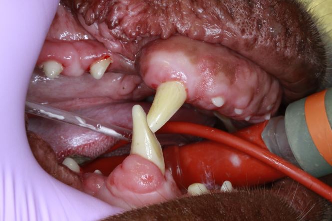
Fig 1 Photograph showing class 1 malocclusion and abnormal wear pattern affecting the maxillary and mandibular canine teeth.
Fig 2 Three dimensional CT image showing proliferative new bone affecting the right coronoid process and zygomatic arch
Fig 3 Transverse CT image showing evidence of fragmentation of the retroarticular process of the right TMJ
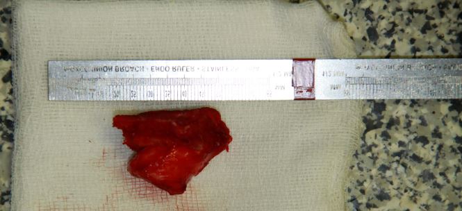
Fig 4 Photograph showing the excised portion of the right zygomatic arch including the abnormal proliferative bone

by Peter Southerden
Both Peter Southerden and Andrew Perry are happy to discuss or give advice on all matters relating to small animal veterinary dentistry or oral surgery. Please phone 01793 528341 or email Eastcott Referrals