Home »
Categories
Blog Articles and News
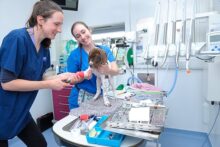
Eastcott completes £3 million expansion
General Blog and News |
We’ve just completed an 18-month, £3 million extension and refurbishment programme at our practice on Dorcan Way which means we can now deliver healthcare and facilities to rival those enjoyed by humans.

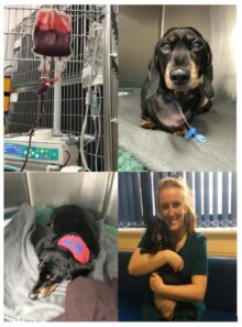
Pet Blood Bank donation session – Wednesday 7th October
General Blog and News |
We’re teaming up with Pet Blood Bank to provide a centre for them to perform regular blood donation sessions.
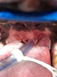
BOAS surgery what’s involved?
Soft Tissue |
Soft Tissue Veterinary Surgeon Hannah Prestwood explains why certain dog breeds have breathing difficulties and what can be done to help them.
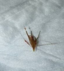
Grass seed danger for dogs
Soft Tissue |
Dry sunny spells see a greater number of seeds falling to the ground, where they can work their way into the hair of a dog’s paw, ears, armpits, tail or skin.
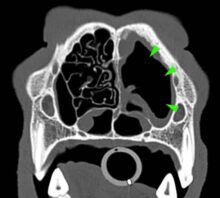
What’s your diagnosis? Computed Tomography Case 6
Diagnostic Imaging |
An 11-year-old cocker spaniel was referred to Eastcott Referrals for a chronic nasal discharge and soft tissue swelling in the dorsal aspect of the nose.
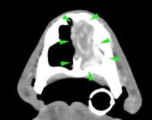
What’s your diagnosis? Computed Tomography Case 5
Diagnostic Imaging |
A 3-year-old Siberian husky was presented for chronic nasal discharge. A CT scan of the head showed marked loss of turbinates in the rostral aspect of both nasal cavities, more severe on the left side (green arrowheads).
Pet Blood Bank donation session – Tuesday 11th August
General Blog and News |
We’re teaming up Pet Blood Bank to provide a centre for them to perform regular blood donation sessions.
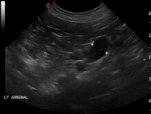
What’s your diagnosis? Computed Tomography Case 4
Diagnostic Imaging |
A 8-year-old cocker spaniel was referred for polyuria/polydipsia and polyphagia. The abdominal ultrasound revealed bilateral symmetric adrenal enlargement.
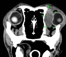
What’s your diagnosis? Computed Tomography Case 3
Diagnostic Imaging |
A 7-years-old Labrador was presented to with lethargy, anorexia and exophthalmos. A CT scan of the head revealed the presence of a soft tissue space-occupying lesion of approximately 3 cm in diameter, arising from the orbital part of the left frontal bone (green arrowheads).
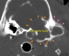
What’s your diagnosis? Computed Tomography Case 2
Diagnostic Imaging |
A 10-year-old Staffordshire bull terrier was referred to Eastcott due to a left sided facial swelling, temporal muscle wastage and difficulty to open his mouth.